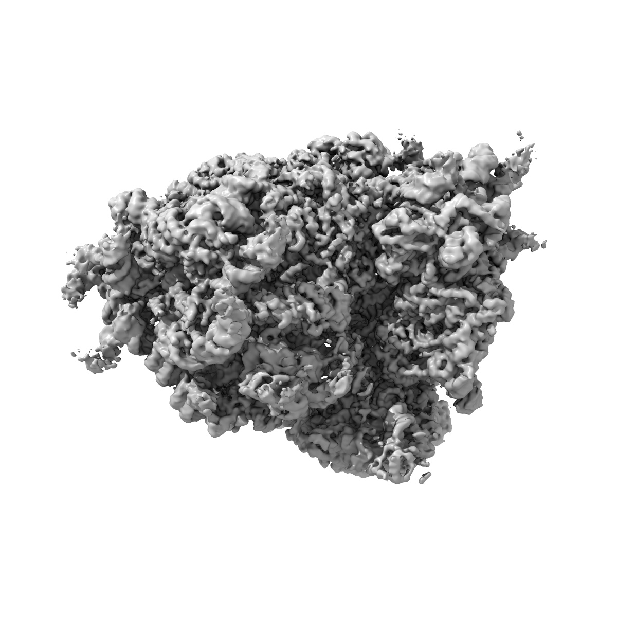
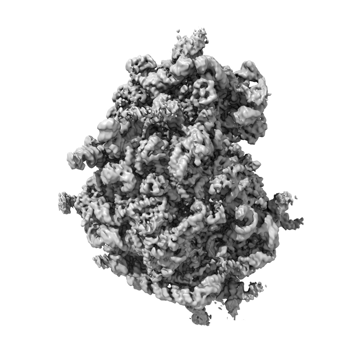
EMD-7834
Mammalian 80S ribosome with a double translocated CrPV-IRES, P-site tRNA and eRF1.
EMD-7834
Single-particle3.2 Å
 Deposition: 27/04/2018
Map released: 06/06/2018
Last modified: 25/12/2024
Sample Organism:
Oryctolagus cuniculus,
Escherichia coli,
Homo sapiens,
Cricket paralysis virus
Sample: Mammalian 80S ribosome in complex with a double translocated CrPV IRES, P-site tRNA and eRF1
Fitted models: 6d90 (Avg. Q-score: 0.335)
Deposition Authors: Pisareva VP, Pisarev AV
Sample: Mammalian 80S ribosome in complex with a double translocated CrPV IRES, P-site tRNA and eRF1
Fitted models: 6d90 (Avg. Q-score: 0.335)
Deposition Authors: Pisareva VP, Pisarev AV
Dual tRNA mimicry in the Cricket Paralysis Virus IRES uncovers an unexpected similarity with the Hepatitis C Virus IRES.

Abstract:
Co-opting the cellular machinery for protein production is a compulsory requirement for viruses. The Cricket Paralysis Virus employs an Internal Ribosomal Entry Site (CrPV-IRES) to express its structural genes in the late stage of infection. Ribosome hijacking is achieved by a sophisticated use of molecular mimicry to tRNA and mRNA, employed to manipulate intrinsically dynamic components of the ribosome. Binding and translocation through the ribosome is required for this IRES to initiate translation. We report two structures, solved by single particle electron cryo-microscopy (cryoEM), of a double translocated CrPV-IRES with aminoacyl-tRNA in the peptidyl site (P site) of the ribosome. CrPV-IRES adopts a previously unseen conformation, mimicking the acceptor stem of a canonical E site tRNA. The structures suggest a mechanism for the positioning of the first aminoacyl-tRNA shared with the distantly related Hepatitis C Virus IRES.
Co-opting the cellular machinery for protein production is a compulsory requirement for viruses. The Cricket Paralysis Virus employs an Internal Ribosomal Entry Site (CrPV-IRES) to express its structural genes in the late stage of infection. Ribosome hijacking is achieved by a sophisticated use of molecular mimicry to tRNA and mRNA, employed to manipulate intrinsically dynamic components of the ribosome. Binding and translocation through the ribosome is required for this IRES to initiate translation. We report two structures, solved by single particle electron cryo-microscopy (cryoEM), of a double translocated CrPV-IRES with aminoacyl-tRNA in the peptidyl site (P site) of the ribosome. CrPV-IRES adopts a previously unseen conformation, mimicking the acceptor stem of a canonical E site tRNA. The structures suggest a mechanism for the positioning of the first aminoacyl-tRNA shared with the distantly related Hepatitis C Virus IRES.