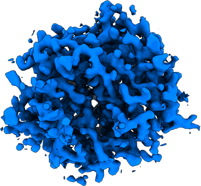
MicroED structure of bovine liver catalase with missing cone solved by suspended drop
Resolution: 4.0 Å
EM Method: Electron Crystallography
Fitted PDBs: 9bdj
Q-score: 0.483
Gillman C, Bu G, Danelius E, Hattne J, Nannenga BL, Gonen T
J Struct Biol X (2024) 9 pp. 100102-100102 [ DOI: doi:10.1016/j.yjsbx.2024.100102 Pubmed: 38962493 ]
- Nadph dihydro-nicotinamide-adenine-dinucleotide phosphate (745 Da, Ligand)
- Bovine liver catalase (Complex from Bos taurus)
- Catalase (59 kDa, Protein from Bos taurus)
- Protoporphyrin ix containing fe (616 Da, Ligand)
MicroED structure of the C11 cysteine protease clostripain
Ruma YN, Bu G, Hattne J, Gonen T
J Struct Biol X (2024) 10 pp. 100107-100107 [ DOI: doi:10.1016/j.yjsbx.2024.100107 Pubmed: 39100863 ]
- Clostripain (59 kDa, Complex from Hathewaya histolytica)
- Sodium ion (22 Da, Ligand)
- Water (18 Da, Ligand)
- Clostripain (59 kDa, Protein from Hathewaya histolytica)
Resolution: 2.15 Å
EM Method: Electron Crystallography
Fitted PDBs: 8vd7
Q-score: 0.717
Gillman C, Bu G, Danelius E, Hattne J, Nannenga BL, Gonen T
J Struct Biol X (2024) 9 pp. 100102-100102 [ DOI: doi:10.1016/j.yjsbx.2024.100102 Pubmed: 38962493 ]
- Water (18 Da, Ligand)
- Chloride ion (35 Da, Ligand)
- 3c-like proteinase nsp5 (33 kDa, Protein from Severe acute respiratory syndrome coronavirus 2)
- Sars-cov-2 main protease (mpro) (Complex from Severe acute respiratory syndrome coronavirus 2)
MicroED structure of an Aeropyrum pernix protoglobin mutant
Resolution: 2.1 Å
EM Method: Electron Crystallography
Fitted PDBs: 8eum
Q-score: 0.741
Danelius E, Porter NJ, Unge J, Arnold FH, Gonen T
J Am Chem Soc (2023) 145 pp. 7159-7165 [ DOI: doi:10.1021/jacs.2c12004 Pubmed: 36948184 ]
- Homotetramer of aeropyrum pernix protoglobin (apepgb) c45g, w59l, y60v, v63r, c102s, f145q, i149l mutant (Complex from Aeropyrum pernix)
- Imidazole (69 Da, Ligand)
- Protoporphyrin ix containing fe (616 Da, Ligand)
- Protogloblin appgb (22 kDa, Protein from Aeropyrum pernix)
- Water (18 Da, Ligand)
- Fe (iii) ion (55 Da, Ligand)
The MicroED structure of proteinase K crystallized by suspended drop crystallization
Resolution: 2.1 Å
EM Method: Electron Crystallography
Fitted PDBs: 8sdk
Q-score: 0.739
Gillman C, Nicolas WJ, Martynowycz MW, Gonen T
IUCrJ (2023) 10 pp. 430-436 [ DOI: doi:10.1107/S2052252523004141 Pubmed: 37223996 ]
- Calcium ion (40 Da, Ligand)
- Proteinase k from tritirachium album (28 kDa, Cellular component from Parengyodontium album)
- Sulfate ion (96 Da, Ligand)
- Proteinase k (28 kDa, Protein from Parengyodontium album)
- Water (18 Da, Ligand)
MicroED structure of Proteinase K from lamellae milled from multiple plasma sources
Resolution: 1.39 Å
EM Method: Electron Crystallography
Fitted PDBs: 8fyo
Q-score: 0.911
Martynowycz MW, Shiriaeva A, Clabbers MTB, Nicolas WJ, Weaver SJ, Hattne J, Gonen T
Nat Commun (2023) 14 pp. 1086-1086 [ Pubmed: 36841804 DOI: doi:10.1038/s41467-023-36733-4 ]
- Calcium ion (40 Da, Ligand)
- Proteinase k (28 kDa, Protein from Parengyodontium album)
- Proteinase k (28 kDa, Complex from Parengyodontium album)
- Nitrate ion (62 Da, Ligand)
- Water (18 Da, Ligand)
MicroED structure of an Aeropyrum pernix protoglobin metallo-carbene complex
Danelius E, Porter NJ, Unge J, Arnold FH, Gonen T
J Am Chem Soc (2023) 145 pp. 7159-7165 [ DOI: doi:10.1021/jacs.2c12004 Pubmed: 36948184 ]
- Protogloblin appgb (22 kDa, Protein from Aeropyrum pernix)
- Homodimer of aeropyrum pernix protoglobin (apepgb) c45g, w59l, y60v, v63r, c102s, f145q, i149l mutant (Complex from Aeropyrum pernix)
- Water (18 Da, Ligand)
- Benzyl[3,3'-(7,12-diethenyl-3,8,13,17-tetramethylporphyrin-2,18-diyl-kappa~4~n~21~,n~22~,n~23~,n~24~)di(propanoato)(2-)]iron (707 Da, Ligand)
MicroED structure of triclinic lysozyme
Clabbers MTB, Martynowycz MW, Hattne J, Gonen T
J Struct Biol X (2022) 6 pp. 100078-100078 [ DOI: doi:10.1016/j.yjsbx.2022.100078 Pubmed: 36507068 ]
- Lysozyme c (14 kDa, Protein from Gallus gallus)
- Nitrate ion (62 Da, Ligand)
- Water (18 Da, Ligand)
- Lysozyme (14 kDa, Complex from Gallus gallus)
MicroED structure of A2A from plasma milled lamellae
Resolution: 2.0 Å
EM Method: Electron Crystallography
Fitted PDBs: 8fyn
Q-score: 0.711
Martynowycz MW, Shiriaeva A, Clabbers MTB, Nicolas WJ, Weaver SJ, Hattne J, Gonen T
Nat Commun (2023) 14 pp. 1086-1086 [ Pubmed: 36841804 DOI: doi:10.1038/s41467-023-36733-4 ]
- (2s)-2,3-dihydroxypropyl (9z)-octadec-9-enoate (356 Da, Ligand)
- Adenosine receptor a2a,soluble cytochrome b562 (49 kDa, Protein from Homo sapiens)
- Oleic acid (282 Da, Ligand)
- Sodium ion (22 Da, Ligand)
- A2a bril adenosine receptor (41 kDa, Complex from Homo sapiens)
- 4-{2-[(7-amino-2-furan-2-yl[1,2,4]triazolo[1,5-a][1,3,5]triazin-5-yl)amino]ethyl}phenol (337 Da, Ligand)
- Cholesterol (386 Da, Ligand)
- Water (18 Da, Ligand)
- (2r)-2,3-dihydroxypropyl (9z)-octadec-9-enoate (356 Da, Ligand)
MicroED structure of Proteinase K from xenon milled lamellae
Resolution: 1.45 Å
EM Method: Electron Crystallography
Fitted PDBs: 8fyp
Q-score: 0.898
Martynowycz MW, Shiriaeva A, Clabbers MTB, Nicolas WJ, Weaver SJ, Hattne J, Gonen T
Nat Commun (2023) 14 pp. 1086-1086 [ Pubmed: 36841804 DOI: doi:10.1038/s41467-023-36733-4 ]
- Calcium ion (40 Da, Ligand)
- Proteinase k (28 kDa, Protein from Parengyodontium album)
- Proteinase k (28 kDa, Complex from Parengyodontium album)
- Nitrate ion (62 Da, Ligand)
- Water (18 Da, Ligand)


