
Negatively stained reconstruction of a rubisco activase
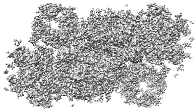
Resolution: 3.02 Å
EM Method: Single-particle
Fitted PDBs: 6jlu
Q-score: 0.385
Pi X, Zhao S, Wang W, Liu D, Xu C, Han G, Kuang T, Sui SF, Shen JR
Science (2019) 365 [ Pubmed: 31371578 DOI: doi:10.1126/science.aax4406 ]
- Extrinsic protein in photosystem ii (10 kDa, Protein from Chaetoceros gracilis)
- Psbg (6 kDa, Protein from Chaetoceros gracilis)
- Psbj (3 kDa, Protein from Chaetoceros gracilis)
- Psii-fcp supercomplex (Complex from Chaetoceros gracilis)
- Psbk (4 kDa, Protein from Chaetoceros gracilis)
- 2,3-dimethyl-5-(3,7,11,15,19,23,27,31,35-nonamethyl-2,6,10,14,18,22,26,30,34-hexatriacontanonaenyl-2,5-cyclohexadiene-1,4-dione-2,3-dimethyl-5-solanesyl-1,4-benzoquinone (749 Da, Ligand)
- Psb34 (2 kDa, Protein from Chaetoceros gracilis)
- Beta-carotene (536 Da, Ligand)
- Chlorophyll c1 (610 Da, Ligand)
- Psbw (5 kDa, Protein from Chaetoceros gracilis)
- Photosystem ii reaction center protein h (7 kDa, Protein from Chaetoceros gracilis)
- Bicarbonate ion (61 Da, Ligand)
- Psbm (4 kDa, Protein from Chaetoceros gracilis)
- Fe (ii) ion (55 Da, Ligand)
- Psbe (9 kDa, Protein from Chaetoceros gracilis)
- Psbt (3 kDa, Protein from Chaetoceros gracilis)
- 1,2-dipalmitoyl-phosphatidyl-glycerole (722 Da, Ligand)
- (3s,3's,5r,5'r,6s,6'r,8'r)-3,5'-dihydroxy-8-oxo-6',7'-didehydro-5,5',6,6',7,8-hexahydro-5,6-epoxy-beta,beta-caroten-3'-yl acetate (658 Da, Ligand)
- Chlorophyll c2 (608 Da, Ligand)
- Water (18 Da, Ligand)
- Cytochrome c-550 (14 kDa, Protein from Chaetoceros gracilis)
- Chloride ion (35 Da, Ligand)
- Extrinsic protein in photosystem ii (26 kDa, Protein from Chaetoceros gracilis)
- 1,2-di-o-acyl-3-o-[6-deoxy-6-sulfo-alpha-d-glucopyranosyl]-sn-glycerol (795 Da, Ligand)
- (3s,3'r,5r,6s,7cis)-7',8'-didehydro-5,6-dihydro-5,6-epoxy-beta,beta-carotene-3,3'-diol (582 Da, Ligand)
- Pheophytin a (871 Da, Ligand)
- Psbl (4 kDa, Protein from Chaetoceros gracilis)
- 1,2-distearoyl-monogalactosyl-diglyceride (787 Da, Ligand)
- Extrinsic protein in photosystem ii (22 kDa, Protein from Chaetoceros gracilis)
- Psbf (3 kDa, Protein from Chaetoceros gracilis)
- Psbi (3 kDa, Protein from Chaetoceros gracilis)
- Fcp-a (18 kDa, Protein from Chaetoceros gracilis)
- Psbb (56 kDa, Protein from Chaetoceros gracilis)
- Fcp-e (18 kDa, Protein from Chaetoceros gracilis)
- Psb31 (18 kDa, Protein from Chaetoceros gracilis)
- Psby (3 kDa, Protein from Chaetoceros gracilis)
- Photosystem ii reaction center x protein (3 kDa, Protein from Chaetoceros gracilis)
- Protoporphyrin ix containing fe (616 Da, Ligand)
- Psba (39 kDa, Protein from Chaetoceros gracilis)
- Psbd (37 kDa, Protein from Chaetoceros gracilis)
- Psbc (49 kDa, Protein from Chaetoceros gracilis)
- Ca-mn4-o5 cluster (339 Da, Ligand)
- Chlorophyll a (893 Da, Ligand)
- Digalactosyl diacyl glycerol (dgdg) (949 Da, Ligand)
- Fcp-f (18 kDa, Protein from Chaetoceros gracilis)
- Psbz (6 kDa, Protein from Chaetoceros gracilis)
- Fcp-d (24 kDa, Protein from Chaetoceros gracilis)
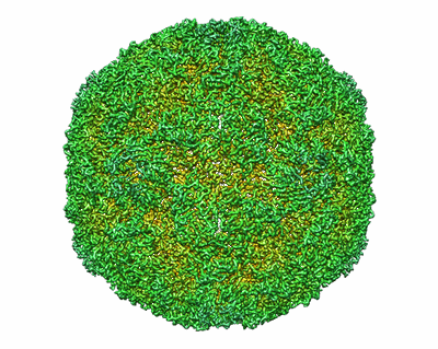
The structure of Enterovirus D68 mature virion
Resolution: 3.4 Å
EM Method: Single-particle
Fitted PDBs: 6aj0
Q-score: 0.547
Zheng Q, Zhu R, Xu L, He M, Yan X, Liu D, Yin Z, Wu Y, Li Y, Yang L, Hou W, Li S, Li Z, Chen Z, Li Z, Yu H, Gu Y, Zhang J, Baker TS, Zhou ZH, Graham BS, Cheng T, Li S, Xia N
Nat Microbiol (2019) 4 pp. 124-133 [ Pubmed: 30397341 DOI: doi:10.1038/s41564-018-0275-7 ]
- Capsid protein vp4 (7 kDa, Protein from Enterovirus D68)
- Virion protein 2 (27 kDa, Protein from Enterovirus D68)
- Capsid protein vp3 (27 kDa, Protein from Enterovirus D68)
- Viral protein 1 (32 kDa, Protein from Enterovirus D68)
- Coxsackievirus a10 (Virus from Coxsackievirus A10)
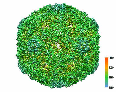
The structure of ICAM-5 triggered Enterovirus D68 virus A-particle
Resolution: 4.0 Å
EM Method: Single-particle
Fitted PDBs: 6aj2
Q-score: 0.412
Zheng Q, Zhu R, Xu L, He M, Yan X, Liu D, Yin Z, Wu Y, Li Y, Yang L, Hou W, Li S, Li Z, Chen Z, Li Z, Yu H, Gu Y, Zhang J, Baker TS, Zhou ZH, Graham BS, Cheng T, Li S, Xia N
Nat Microbiol (2019) 4 pp. 124-133 [ Pubmed: 30397341 DOI: doi:10.1038/s41564-018-0275-7 ]
- Capsid protein vp3 (27 kDa, Protein from Enterovirus D68)
- Capsid protein vp1 (32 kDa, Protein from Enterovirus D68)
- Capsid protein vp2 (27 kDa, Protein from Enterovirus D68)
- Enterovirus d (Virus from Enterovirus D)
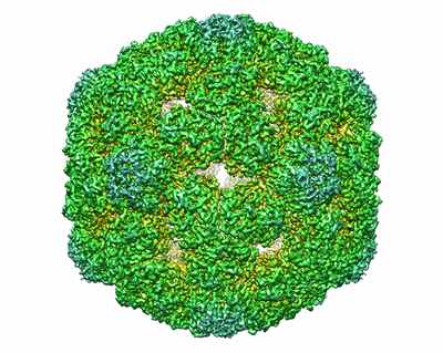
The structure of Enterovirus D68 procapsid
Resolution: 3.8 Å
EM Method: Single-particle
Fitted PDBs: 6aj3
Q-score: 0.472
Zheng Q, Zhu R, Xu L, He M, Yan X, Liu D, Yin Z, Wu Y, Li Y, Yang L, Hou W, Li S, Li Z, Chen Z, Li Z, Yu H, Gu Y, Zhang J, Baker TS, Zhou ZH, Graham BS, Cheng T, Li S, Xia N
Nat Microbiol (2019) 4 pp. 124-133 [ Pubmed: 30397341 DOI: doi:10.1038/s41564-018-0275-7 ]
- Enterovirus d (Virus from Enterovirus d)
- Capsid protein vp3 (27 kDa, Protein from Enterovirus D68)
- Capsid protein vp2 (27 kDa, Protein from Enterovirus D68)
- Capsid protein vp1 (32 kDa, Protein from Enterovirus D68)

The structure of Enterovirus D68 mature virion in complex with Fab 15C5
Resolution: 3.6 Å
EM Method: Single-particle
Fitted PDBs: 6aj7
Q-score: 0.518
Zheng Q, Zhu R, Xu L, He M, Yan X, Liu D, Yin Z, Wu Y, Li Y, Yang L, Hou W, Li S, Li Z, Chen Z, Li Z, Yu H, Gu Y, Zhang J, Baker TS, Zhou ZH, Graham BS, Cheng T, Li S, Xia N
Nat Microbiol (2019) 4 pp. 124-133 [ Pubmed: 30397341 DOI: doi:10.1038/s41564-018-0275-7 ]
- Vl of fab 15c5 (11 kDa, Protein from Mus musculus)
- Capsid protein vp3 (27 kDa, Protein from Enterovirus D68)
- Vh of fab 15c5 (13 kDa, Protein from Mus musculus)
- Enterovirus d68 (Virus from Enterovirus D68)
- Capsid protein vp2 (27 kDa, Protein from Enterovirus D68)
- Capsid protein vp1 (32 kDa, Protein from Enterovirus D68)

The structure of Enterovirus D68 mature virion in complex with Fab 15C5 and 11G1
Resolution: 3.5 Å
EM Method: Single-particle
Fitted PDBs: 6aj9
Q-score: 0.497
Zheng Q, Zhu R, Xu L, He M, Yan X, Liu D, Yin Z, Wu Y, Li Y, Yang L, Hou W, Li S, Li Z, Chen Z, Li Z, Yu H, Gu Y, Zhang J, Baker TS, Zhou ZH, Graham BS, Cheng T, Li S, Xia N
Nat Microbiol (2019) 4 pp. 124-133 [ Pubmed: 30397341 DOI: doi:10.1038/s41564-018-0275-7 ]
- Vl of fab 15c5 (11 kDa, Protein from Mus musculus)
- Vh of fab 11g1 (13 kDa, Protein from Mus musculus)
- Vl of fab 11g1 (12 kDa, Protein from Mus musculus)
- Capsid protein vp3 (27 kDa, Protein from Enterovirus D68)
- Vh of fab 15c5 (13 kDa, Protein from Mus musculus)
- Enterovirus d68 (Virus from Enterovirus D68)
- Capsid protein vp2 (27 kDa, Protein from Enterovirus D68)
- Capsid protein vp1 (32 kDa, Protein from Enterovirus D68)

The structure of Enterovirus D68 A-particle triggered by an unknown stimulus
Zheng Q, Zhu R, Xu L, He M, Yan X, Liu D, Yin Z, Wu Y, Li Y, Yang L, Hou W, Li S, Li Z, Chen Z, Li Z, Yu H, Gu Y, Zhang J, Baker TS, Zhou ZH, Graham BS, Cheng T, Li S, Xia N
Nat Microbiol (2019) 4 pp. 124-133 [ Pubmed: 30397341 DOI: doi:10.1038/s41564-018-0275-7 ]
- Vp3 (Macromolecule from Enterovirus D68)
- Enterovirus d68 (Virus from Enterovirus D68)
- Vp1 (Macromolecule from Enterovirus D68)
- Vp2 (Macromolecule from Enterovirus D68)

Cryo-EM structure of 8nm repeat tubulin lattice of the ciliary microtubule doublet
Resolution: 5.7 Å
EM Method: Single-particle
Fitted PDBs: 5ubq
Q-score: 0.144
Ichikawa M, Liu D, Kastritis PL, Basu K, Hsu TC, Yang S, Bui KH
Nat Commun (2017) 8 pp. 15035-15035 [ DOI: doi:10.1038/ncomms15035 Pubmed: 28462916 ]
- Guanosine-5'-diphosphate (443 Da, Ligand)
- Guanosine-5'-triphosphate (523 Da, Ligand)
- Tubulin alpha chain (48 kDa, Protein from Tetrahymena thermophila)
- Magnesium ion (24 Da, Ligand)
- 8nm-repeat tubulin lattice of microtubule doublet (Cellular component from Tetrahymena thermophila)
- Tubulin beta chain (48 kDa, Protein from Tetrahymena thermophila)

Cryo-EM structure of 16nm repeat ciliary microtubule doublet
Ichikawa M, Liu D, Kastritis PL, Basu K, Hsu TC, Yang S, Bui KH
Nat Commun (2017) 8 pp. 15035-15035 [ DOI: doi:10.1038/ncomms15035 Pubmed: 28462916 ]