
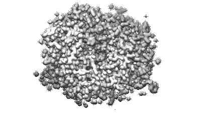
MicroED structure of orthorhombic Vancomycin at 1.2 A resolution
Resolution: 1.2 Å
EM Method: Electron Crystallography
Fitted PDBs: 7c4u
Q-score: 0.803
Fan Q, Li L, Xue H, Zhou H, Zhao L, Liu J, Mao J, Wu S, Zhang S, Wu C, Li X, Zhou X, Wang J
Angew Chem Int Ed Engl (2020) 59 pp. 15141-15146 [ Pubmed: 32432368 DOI: doi:10.1002/anie.202003922 ]
- Chloride ion (35 Da, Ligand)
- Vancomycin (Complex from Amycolatopsis orientalis)
- Water (18 Da, Ligand)
- Vancomycin (1 kDa, Protein from Amycolatopsis orientalis)

MicroED structure of anorthic Vancomycin at 1.05 A resolution
Resolution: 1.05 Å
EM Method: Electron Crystallography
Fitted PDBs: 7c4v
Q-score: 0.676
Fan Q, Li L, Xue H, Zhou H, Zhao L, Liu J, Mao J, Wu S, Zhang S, Wu C, Li X, Zhou X, Wang J
Angew Chem Int Ed Engl (2020) 59 pp. 15141-15146 [ Pubmed: 32432368 DOI: doi:10.1002/anie.202003922 ]
- Chloride ion (35 Da, Ligand)
- Vancomycin (Complex from Amycolatopsis orientalis)
- Water (18 Da, Ligand)
- Vancomycin (1 kDa, Protein from Amycolatopsis orientalis)
Resolution: 1.5 Å
EM Method: Electron Crystallography
Fitted PDBs: 6law
Q-score: 0.894
J Struct Biol (2019) 205 pp. 59-64 [ DOI: doi:10.1016/j.jsb.2019.02.004 Pubmed: 30794865 ]
- Water (18 Da, Ligand)
- Proteinase k (28 kDa, Protein from Parengyodontium album)
- Proteinase k (Complex from Parengyodontium album)
- Sulfate ion (96 Da, Ligand)

Resolution: 1.73 Å
EM Method: Electron Crystallography
Fitted PDBs: 6lav
Q-score: 0.811
J Struct Biol (2019) 205 pp. 59-64 [ DOI: doi:10.1016/j.jsb.2019.02.004 Pubmed: 30794865 ]
- Water (18 Da, Ligand)
- Lysozyme (Complex from Gallus gallu)
- Lysozyme c (14 kDa, Protein from Gallus gallus)
- Acetate ion (59 Da, Ligand)

200kV MicroED structure of FUS (37-42) SYSGYS solved from merged datasets at 0.65 A
Resolution: 0.65 Å
EM Method: Electron Crystallography
Fitted PDBs: 6kj1
Q-score: 0.973
Zhou H, Luo F, Luo Z, Li D, Liu C, Li X
Anal Chem (2019) 91 pp. 10996-11003 [ DOI: doi:10.1021/acs.analchem.9b01162 Pubmed: 31334636 ]
- Water (18 Da, Ligand)
- Fus lc rac1 (Complex)
- Rna-binding protein fus (662 Da, Protein from Homo sapiens)

200kV MicroED structure of FUS (37-42) SYSGYS solved from single crystal at 0.67 A
Resolution: 0.67 Å
EM Method: Electron Crystallography
Fitted PDBs: 6kj2
Q-score: 0.972
Zhou H, Luo F, Luo Z, Li D, Liu C, Li X
Anal Chem (2019) 91 pp. 10996-11003 [ DOI: doi:10.1021/acs.analchem.9b01162 Pubmed: 31334636 ]
- Water (18 Da, Ligand)
- Fus lc rac1 (Complex)
- Rna-binding protein fus (662 Da, Protein from Homo sapiens)

120kV MicroED structure of FUS (37-42) SYSGYS solved from merged datasets at 0.60 A
Resolution: 0.6 Å
EM Method: Electron Crystallography
Fitted PDBs: 6kj3
Q-score: 0.962
Zhou H, Luo F, Luo Z, Li D, Liu C, Li X
Anal Chem (2019) 91 pp. 10996-11003 [ DOI: doi:10.1021/acs.analchem.9b01162 Pubmed: 31334636 ]
- Water (18 Da, Ligand)
- Fus lc rac1 (Complex)
- Rna-binding protein fus (662 Da, Protein from Homo sapiens)

120kV MicroED structure of FUS (37-42) SYSGYS solved from single crystal at 0.65 A
Resolution: 0.65 Å
EM Method: Electron Crystallography
Fitted PDBs: 6kj4
Q-score: 0.963
Zhou H, Luo F, Luo Z, Li D, Liu C, Li X
Anal Chem (2019) 91 pp. 10996-11003 [ DOI: doi:10.1021/acs.analchem.9b01162 Pubmed: 31334636 ]
- Water (18 Da, Ligand)
- Fus lc rac1 (Complex)
- Rna-binding protein fus (662 Da, Protein from Homo sapiens)