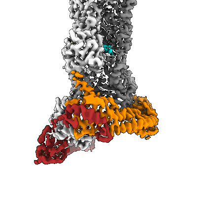
Structure of the formin Cdc12 bound to the barbed end of phalloidin-stabilized F-actin.
Resolution: 3.56 Å
EM Method: Single-particle
Fitted PDBs: 8rtt
Q-score: 0.444
Oosterheert W, Boiero Sanders M, Funk J, Prumbaum D, Raunser S, Bieling P
Science (2024) 384 pp. eadn9560-eadn9560 [ Pubmed: 38603491 DOI: doi:10.1126/science.adn9560 ]
- Phalloidin. (Complex from Amanita phalloides)
- Magnesium ion (24 Da, Ligand)
- Actin filament (Complex from Homo sapiens)
- Cell division control protein 12 (48 kDa, Protein from Schizosaccharomyces pombe)
- Actin, cytoplasmic 1, n-terminally processed (41 kDa, Protein from Homo sapiens)
- Adenosine-5'-diphosphate (427 Da, Ligand)
- Phosphate ion (94 Da, Ligand)
- Complex of the dimeric fh2 domain of s. pombe cdc12 bound to the barbed end of phalloidin stabilized f-actin. (Complex)
- Phalloidin (808 Da, Protein from Amanita phalloides)
- Dimeric fh2 domain of s. pombe cdc12 (Complex from Schizosaccharomyces pombe)
Resolution: 6.25 Å
EM Method: Single-particle
Fitted PDBs: 8rty
Q-score: 0.17
Oosterheert W, Boiero Sanders M, Funk J, Prumbaum D, Raunser S, Bieling P
Science (2024) 384 pp. eadn9560-eadn9560 [ Pubmed: 38603491 DOI: doi:10.1126/science.adn9560 ]
- Dimeric fh1fh2 domain of s. pombe cdc12 (Complex from Schizosaccharomyces pombe)
- Magnesium ion (24 Da, Ligand)
- Actin filament (Complex from Homo sapiens)
- Adenosine-5'-diphosphate (427 Da, Ligand)
- Methylated-dna--protein-cysteine methyltransferase,cell division control protein 12 (77 kDa, Protein from Schizosaccharomyces pombe)
- Phosphate ion (94 Da, Ligand)
- Profilin-1 (15 kDa, Protein from Homo sapiens)
- Actin, cytoplasmic 1, n-terminally processed (41 kDa, Protein from Homo sapiens)
- Profilin-1-s29c/s71m (Complex from Homo sapiens)
- Phalloidin (amanita phalloides) (808 Da, Protein from Amanita phalloides)
- Phalloidin (Complex from Amanita phalloides)
- Actin-formin-profilin ring complex: the phalloidin-stabilized f-actin barbed end bound by dimeric-cdc12 and profilin-s71m. (Complex)
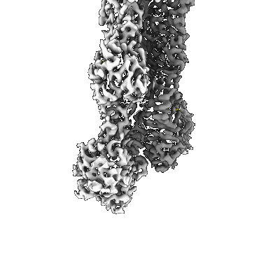
Structure of the undecorated barbed end of F-actin.
Resolution: 3.08 Å
EM Method: Single-particle
Fitted PDBs: 8ru0
Q-score: 0.542
Oosterheert W, Boiero Sanders M, Funk J, Prumbaum D, Raunser S, Bieling P
Science (2024) 384 pp. eadn9560-eadn9560 [ Pubmed: 38603491 DOI: doi:10.1126/science.adn9560 ]
- Magnesium ion (24 Da, Ligand)
- Actin filament (Complex)
- Adenosine-5'-diphosphate (427 Da, Ligand)
- Complex of the actin subunits that form the barbed end of actin filaments. (Complex from Oryctolagus cuniculus)
- Adenosine-5'-triphosphate (507 Da, Ligand)
- Actin, alpha skeletal muscle (41 kDa, Protein from Oryctolagus cuniculus)
- Phosphate ion (94 Da, Ligand)
Structure of the F-actin barbed end bound by formin mDia1
Resolution: 3.49 Å
EM Method: Single-particle
Fitted PDBs: 8ru2
Q-score: 0.362
Oosterheert W, Boiero Sanders M, Funk J, Prumbaum D, Raunser S, Bieling P
Science (2024) 384 pp. eadn9560-eadn9560 [ DOI: doi:10.1126/science.adn9560 Pubmed: 38603491 ]
- Actin, cytoplasmic 1, n-terminally processed (41 kDa, Protein from Homo sapiens)
- Magnesium ion (24 Da, Ligand)
- Methylated-dna--protein-cysteine methyltransferase,protein diaphanous homolog 1 (86 kDa, Protein from Mus musculus)
- Actin filament (Complex from Homo sapiens)
- Mdia1-bound f-actin barbed end. (Complex)
- Mouse mdia1 (fh1fh2c domain) (Complex from Mus musculus)
- Adenosine-5'-diphosphate (427 Da, Ligand)
Structure of the formin INF2 bound to the barbed end of F-actin.
Resolution: 3.41 Å
EM Method: Single-particle
Fitted PDBs: 8rv2
Q-score: 0.43
Oosterheert W, Boiero Sanders M, Funk J, Prumbaum D, Raunser S, Bieling P
Science (2024) 384 pp. eadn9560-eadn9560 [ Pubmed: 38603491 DOI: doi:10.1126/science.adn9560 ]
- Inf2 dimer (Complex from Homo sapiens)
- Magnesium ion (24 Da, Ligand)
- Adenosine-5'-diphosphate (427 Da, Ligand)
- Adenosine-5'-triphosphate (507 Da, Ligand)
- Complex of the formin inf2 dimer that binds to the actin subunits at the barbed end of actin filaments. (Complex)
- Actin, alpha skeletal muscle (41 kDa, Protein from Oryctolagus cuniculus)
- Isoform 2 of inverted formin-2 (84 kDa, Protein from Homo sapiens)
- Phosphate ion (94 Da, Ligand)
- Actin filament barbed end (Complex from Oryctolagus cuniculus)
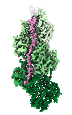
In situ structure of nebulin bound to actin filament in skeletal sarcomere
Resolution: 4.5 Å
EM Method: Subtomogram averaging
Fitted PDBs: 7qim
Q-score: 0.312
Wang Z, Grange M, Pospich S, Wagner T, Kho AL, Gautel M, Raunser S
Science (2022) 375 pp. eabn1934-eabn1934 [ Pubmed: 35175800 DOI: doi:10.1126/science.abn1934 ]
- Mouse psoas muscle myofibrils (Cellular component from Mus musculus)
- Magnesium ion (24 Da, Ligand)
- Nebulin (mouse) (6 kDa, Protein from Mus musculus)
- Adenosine-5'-diphosphate (427 Da, Ligand)
- Acts protein (42 kDa, Protein from house mouse)
- Nebulin (mouse) (9 kDa, Protein from Mus musculus)
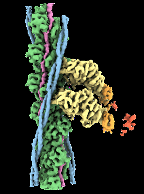
In situ structure of actomyosin complex in skeletal sarcomere
Resolution: 6.6 Å
EM Method: Subtomogram averaging
Fitted PDBs: 7qin
Q-score: 0.196
Wang Z, Grange M, Pospich S, Wagner T, Kho AL, Gautel M, Raunser S
Science (2022) 375 pp. eabn1934-eabn1934 [ Pubmed: 35175800 DOI: doi:10.1126/science.abn1934 ]
- Mouse psoas muscle myofibrils (Cellular component from Mus musculus)
- Magnesium ion (24 Da, Ligand)
- Nebulin (mouse) (9 kDa, Protein from house mouse)
- Actin, alpha skeletal muscle (41 kDa, Protein from house mouse)
- Tropomyosin, alpha-1 (mouse) (10 kDa, Protein from house mouse)
- Adenosine-5'-diphosphate (427 Da, Ligand)
- Myosin-4 (96 kDa, Protein from house mouse)
- Tropomyosin, alpha-1 (mouse) (7 kDa, Protein from house mouse)
- Nebulin (mouse) (6 kDa, Protein from house mouse)
- Tropomyosin, alpha-1 (mouse) (9 kDa, Protein from house mouse)
In situ structure of myosin neck domain in skeletal sarcomere (centered on regulatory light chain)
Resolution: 9.0 Å
EM Method: Subtomogram averaging
Fitted PDBs: 7qio
Q-score: 0.089
Wang Z, Grange M, Pospich S, Wagner T, Kho AL, Gautel M, Raunser S
Science (2022) 375 pp. eabn1934-eabn1934 [ Pubmed: 35175800 DOI: doi:10.1126/science.abn1934 ]
- Myosin-4 (96 kDa, Protein from Mus musculus)
- Myosin regulatory light chain 2, skeletal muscle isoform (18 kDa, Protein from Mus musculus)
- Mouse psoas muscle myofibrils (Cellular component from Mus musculus)
- Myosin light chain 1/3, skeletal muscle isoform (20 kDa, Protein from Mus musculus)


