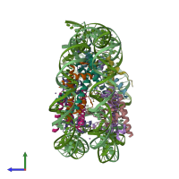
5b1l

X-ray diffraction
2.35Å resolution

The mouse nucleosome structure containing H3t

Released:
Model geometry
Fit model/data

Function and Biology Details

Structure analysis Details

Assembly composition:
hetero decamer (preferred)
PDBe Complex ID:
PDB-CPX-110963 (preferred)
Entry contents:
4 distinct polypeptide molecules
1 distinct DNA molecule
Macromolecules (5 distinct):
Histone H3.1 Chains: A, E
Molecule details ›
Chains: A, E
Length: 139 amino acids
Theoretical weight: 15.74 KDa
Source organism: Mus musculus
Expression system: Escherichia coli BL21(DE3)
UniProt:
  • Canonical: P68433 (Residues: 1-136; Coverage: 100%)
Gene names: H3.1-221, H3.1-291, H3.1-I, H3a, H3c1, H3c10, H3c11, H3c8, H3g, H3h, H3i, Hist1h3a, Hist1h3g, Hist1h3h, Hist1h3i
Sequence domains: Core histone H2A/H2B/H3/H4
Structure domains: Histone, subunit A
Histone H4 Chains: B, F
Molecule details ›
Chains: B, F
Length: 106 amino acids
Theoretical weight: 11.68 KDa
Source organism: Mus musculus
Expression system: Escherichia coli
UniProt:
  • Canonical: P62806 (Residues: 1-103; Coverage: 100%)
Gene names: H4-12, H4-53, H4c1, H4c11, H4c12, H4c14, H4c16, H4c2, H4c3, H4c4, H4c6, H4c8, H4c9, H4f16, Hist1h4a, Hist1h4b, Hist1h4c, Hist1h4d, Hist1h4f, Hist1h4h, Hist1h4i, Hist1h4j, Hist1h4k, Hist1h4m, Hist2h4, Hist2h4a, Hist4h4
Sequence domains: Centromere kinetochore component CENP-T histone fold
Structure domains: Histone, subunit A
Histone H2A type 1-B Chains: C, G
H2B.U histone 2 Chains: D, H
DNA (146-MER) Chains: I, J

Ligands and Environments

2 bound ligands:
No modified residues

Experiments and Validation Details

Entry percentile scores
X-ray source: PHOTON FACTORY BEAMLINE BL-1A
Spacegroup: P212121
Unit cell:
a: 98.968Å b: 107.425Å c: 167.332Å
α: 90° β: 90° γ: 90°
R-values:
R R work R free
0.199 0.197 0.243
Expression systems:
  • Escherichia coli BL21(DE3)
  • Escherichia coli