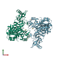
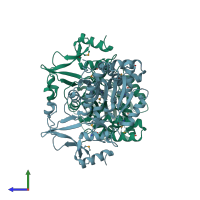
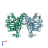
1zkd

X-ray diffraction
2.1Å resolution

X-Ray structure of the putative protein Q6N1P6 from Rhodopseudomonas palustris at the resolution 2.1 A , Northeast Structural Genomics Consortium target RpR58

Released:
Model geometry
Fit model/data
Entry authors: Kuzin AP, Yong W, Vorobiev SM, Acton T, Ma L, Xiao R, Montelione G, Tong L, Hunt JF, Northeast Structural Genomics Consortium (NESG)

Function and Biology Details

Structure analysis Details

Assembly composition:
homo dimer (preferred)
Assembly name:
PDBe Complex ID:
PDB-CPX-179939 (preferred)
Entry contents:
1 distinct polypeptide molecule
Macromolecule:
DUF185 Chains: A, B
Molecule details ›
Chains: A, B
Length: 387 amino acids
Theoretical weight: 42.82 KDa
Source organism: Rhodopseudomonas palustris CGA009
Expression system: Escherichia coli
UniProt:
  • Canonical: Q6N1P6 (Residues: 1-379; Coverage: 100%)
Gene names: RPA4359, TX73_022595
Sequence domains: Putative S-adenosyl-L-methionine-dependent methyltransferase
Structure domains:

Ligands and Environments

No bound ligands
1 modified residue:

Experiments and Validation Details

Entry percentile scores
X-ray source: NSLS BEAMLINE X4A
Spacegroup: P61
Unit cell:
a: 79.795Å b: 79.795Å c: 247.352Å
α: 90° β: 90° γ: 120°
R-values:
R R work R free
0.223 0.223 0.258
Expression system: Escherichia coli