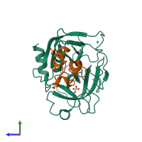
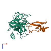
Function and Biology Details
Reaction catalysed:
Preferential cleavage: Arg-|-, Lys-|-.
Biochemical function:
Biological process:
Cellular component:
Sequence domains:
- Pancreatic trypsin inhibitor Kunitz domain
- Peptidase S1A, chymotrypsin family
- Serine proteases, trypsin domain
- Pancreatic trypsin inhibitor Kunitz domain superfamily
- Peptidase S1, PA clan, chymotrypsin-like fold
- Proteinase inhibitor I2, Kunitz, conserved site
- Serine proteases, trypsin family, serine active site
- Serine proteases, trypsin family, histidine active site
1 more domain
Structure domains:
Structure analysis Details
Assemblies composition:
Assembly name:
PDBe Complex ID:
PDB-CPX-133321 (preferred)
Entry contents:
2 distinct polypeptide molecules
Macromolecules (2 distinct):


