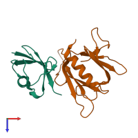
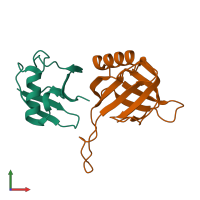
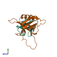
2hth

X-ray diffraction
2.7Å resolution

Structural basis for ubiquitin recognition by the human EAP45/ESCRT-II GLUE domain

Released:

Function and Biology Details

Structure analysis Details

Assembly composition:
hetero dimer (preferred)
PDBe Complex ID:
PDB-CPX-533877 (preferred)
Entry contents:
2 distinct polypeptide molecules
Macromolecules (2 distinct):
Ubiquitin Chain: A
Molecule details ›
Chain: A
Length: 76 amino acids
Theoretical weight: 8.58 KDa
Source organism: Homo sapiens
Expression system: Escherichia coli
UniProt:
  • Canonical: P0CG48 (Residues: 609-684; Coverage: 11%)
Gene name: UBC
Sequence domains: Ubiquitin family
Structure domains: Phosphatidylinositol 3-kinase Catalytic Subunit; Chain A, domain 1
Vacuolar protein-sorting-associated protein 36 Chain: B
Molecule details ›
Chain: B
Length: 140 amino acids
Theoretical weight: 16.26 KDa
Source organism: Homo sapiens
Expression system: Escherichia coli
UniProt:
  • Canonical: Q86VN1 (Residues: 1-138; Coverage: 36%)
Gene names: C13orf9, CGI-145, EAP45, VPS36
Sequence domains: Vacuolar protein sorting protein 36 Vps36
Structure domains: Pleckstrin-homology domain (PH domain)/Phosphotyrosine-binding domain (PTB)

Ligands and Environments

No bound ligands
No modified residues

Experiments and Validation Details

Entry percentile scores
X-ray source: NSLS BEAMLINE X29A
Spacegroup: P42212
Unit cell:
a: 102.727Å b: 102.727Å c: 54.306Å
α: 90° β: 90° γ: 90°
R-values:
R R work R free
0.252 0.248 0.289
Expression system: Escherichia coli