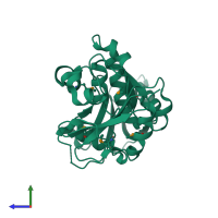
2pgx

X-ray diffraction
2Å resolution

Crystal structure of UPF0341 protein yhiQ from E. coli, Northeast Structural Genomics Target ER585

Released:
Model geometry
Fit model/data
Source organism: Escherichia coli O157:H7
Entry authors: Forouhar F, Su M, Seetharaman J, Janjua H, Fang Y, Xiao R, Liu J, Baran MC, Acton TB, Montelione GT, Tong L, Hunt JF, Northeast Structural Genomics Consortium (NESG)

Function and Biology Details

Reaction catalysed:
S-adenosyl-L-methionine + guanine(1516) in 16S rRNA = S-adenosyl-L-homocysteine + N(2)-methylguanine(1516) in 16S rRNA
Biological process:
Cellular component:

Structure analysis Details

Assembly composition:
monomeric (preferred)
PDBe Complex ID:
PDB-CPX-159483 (preferred)
Entry contents:
1 distinct polypeptide molecule
Macromolecule:
Ribosomal RNA small subunit methyltransferase J Chain: A
Molecule details ›
Chain: A
Length: 258 amino acids
Theoretical weight: 28.34 KDa
Source organism: Escherichia coli O157:H7
Expression system: Escherichia coli
UniProt:
  • Canonical: P68568 (Residues: 1-250; Coverage: 100%)
Gene names: ECs4369, Z4897, rsmJ, yhiQ
Sequence domains: Putative SAM-dependent methyltransferase
Structure domains:

Ligands and Environments

No bound ligands
1 modified residue:

Experiments and Validation Details

Entry percentile scores
X-ray source: NSLS BEAMLINE X4A
Spacegroup: P212121
Unit cell:
a: 40.987Å b: 74.41Å c: 78.122Å
α: 90° β: 90° γ: 90°
R-values:
R R work R free
0.229 0.228 0.243
Expression system: Escherichia coli