2hpv

X-ray diffraction
2Å resolution

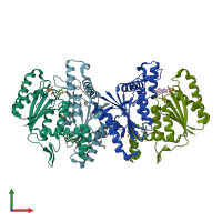
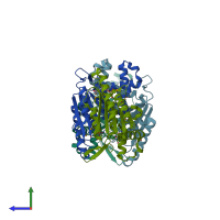
Crystal structure of FMN-Dependent azoreductase from Enterococcus faecalis

Released:
Source organism: Enterococcus faecalis
Entry authors: Liu ZJ, Chen L, Chen H, Rose J, Wang BC, Southeast Collaboratory for Structural Genomics (SECSG)

Function and Biology Details

Reaction catalysed:
Anthranilate + N,N-dimethyl-1,4-phenylenediamine + 2 NAD(+) = 2-(4-dimethylaminophenyl)diazenylbenzoate + 2 NADH
Biological process:
  • not assigned
Cellular component:
  • not assigned

Structure analysis Details

Assembly composition:
homo dimer (preferred)
PDBe Complex ID:
PDB-CPX-182766 (preferred)
Entry contents:
1 distinct polypeptide molecule
Macromolecule:
FMN-dependent NADH:quinone oxidoreductase Chains: A, B, C, D
Molecule details ›
Chains: A, B, C, D
Length: 208 amino acids
Theoretical weight: 23.43 KDa
Source organism: Enterococcus faecalis
Expression system: Escherichia coli
UniProt:
  • Canonical: Q831B2 (Residues: 1-208; Coverage: 100%)
Gene names: EF_2601, azoA, azoR
Sequence domains: Flavodoxin-like fold
Structure domains: Rossmann fold

Ligands and Environments

1 bound ligand:
1 modified residue:

Experiments and Validation Details

Entry percentile scores
X-ray source: APS BEAMLINE 22-ID
Spacegroup: P212121
Unit cell:
a: 98.038Å b: 99.629Å c: 106.737Å
α: 90° β: 90° γ: 90°
R-values:
R R work R free
0.202 0.199 0.25
Expression system: Escherichia coli