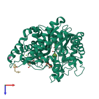
Function and Biology Details
Reactions catalysed:
Hydrolysis of terminal, non-reducing beta-D-glucosyl residues with release of beta-D-glucose
D-glucosyl-N-acylsphingosine + H(2)O = D-glucose + N-acylsphingosine
D-galactosyl-N-acylsphingosine + H(2)O = D-galactose + N-acylsphingosine
Biochemical function:
Biological process:
Cellular component:
Structure analysis Details
Assembly composition:
monomeric (preferred)
Assembly name:
Cytosolic beta-glucosidase (preferred)
PDBe Complex ID:
PDB-CPX-190531 (preferred)
Entry contents:
1 distinct polypeptide molecule
Macromolecule:
Ligands and Environments
Experiments and Validation Details
wwPDB Validation report is not available for this entry.
X-ray source:
SPRING-8 BEAMLINE BL38B1
Spacegroup:
P212121
Expression system: Escherichia coli


