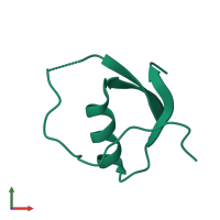
3ovo

X-ray diffraction
1.55Å resolution

REFINED X-RAY CRYSTAL STRUCTURES OF THE REACTIVE SITE MODIFIED OVOMUCOID INHIBITOR THIRD DOMAINS FROM SILVER PHEASANT (OMSVP3(ASTERISK)) AND FROM JAPANESE QUAIL (OMJPQ3(ASTERISK))

Released:

Function and Biology Details

Biological process:
  • not assigned
Cellular component:
  • not assigned

Structure analysis Details

Assembly composition:
monomeric (preferred)
Assembly name:
PDBe Complex ID:
PDB-CPX-133773 (preferred)
Entry contents:
1 distinct polypeptide molecule
Macromolecule:
Ovomucoid Chain: A
Molecule details ›
Chain: A
Length: 56 amino acids
Theoretical weight: 6.07 KDa
Source organism: Coturnix japonica
Expression system: Not provided
UniProt:
  • Canonical: P01003 (Residues: 131-186; Coverage: 30%)
Sequence domains: Kazal-type serine protease inhibitor domain
Structure domains: Wheat Germ Agglutinin (Isolectin 2); domain 1

Ligands and Environments

No bound ligands
No modified residues

Experiments and Validation Details

Entry percentile scores
Spacegroup: P1
Unit cell:
a: 23.55Å b: 21.19Å c: 28.14Å
α: 62.09° β: 115.46° γ: 112.95°
R-values:
R R work R free
0.192 0.192 not available
Expression system: Not provided