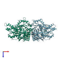
3ua3

X-ray diffraction
3Å resolution

Crystal Structure of Protein Arginine Methyltransferase PRMT5 in complex with SAH

Released:
Model geometry
Fit model/data
Source organism: Caenorhabditis elegans
Primary publication:
Structural insights into protein arginine symmetric dimethylation by PRMT5.
Proc Natl Acad Sci U S A 108 20538-43 (2011)
PMID: 22143770

Function and Biology Details

Reaction catalysed:
(1a) S-adenosyl-L-methionine + [protein]-L-arginine = S-adenosyl-L-homocysteine + [protein]-N(omega)-methyl-L-arginine
Biological process:
Cellular component:

Structure analysis Details

Assembly composition:
homo dimer (preferred)
PDBe Complex ID:
PDB-CPX-155487 (preferred)
Entry contents:
1 distinct polypeptide molecule
Macromolecule:
Protein arginine N-methyltransferase 5 Chains: A, B
Molecule details ›
Chains: A, B
Length: 745 amino acids
Theoretical weight: 85.54 KDa
Source organism: Caenorhabditis elegans
Expression system: Escherichia coli
UniProt:
  • Canonical: P46580 (Residues: 1-734; Coverage: 100%)
Gene names: C34E10.5, prmt-5, tag-251
Sequence domains:
Structure domains:

Ligands and Environments

1 bound ligand:
1 modified residue:

Experiments and Validation Details

Entry percentile scores
X-ray source: SSRF BEAMLINE BL17U
Spacegroup: P212121
Unit cell:
a: 100.376Å b: 129.381Å c: 149.315Å
α: 90° β: 90° γ: 90°
R-values:
R R work R free
0.226 0.223 0.281
Expression system: Escherichia coli