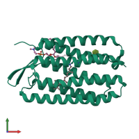
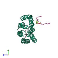
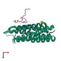
3utw

X-ray diffraction
2.4Å resolution

Crystal structure of bacteriorhodopsin mutant P50A/Y57F

Released:
Primary publication:
Shifting hydrogen bonds may produce flexible transmembrane helices.
Proc Natl Acad Sci U S A 109 8121-6 (2012)
PMID: 22566663

Function and Biology Details

Structure analysis Details

Assemblies composition:
monomeric (preferred)
homo dimer
Assembly name:
PDBe Complex ID:
PDB-CPX-136411 (preferred)
Entry contents:
1 distinct polypeptide molecule
Macromolecule:
Bacteriorhodopsin Chain: A
Molecule details ›
Chain: A
Length: 249 amino acids
Theoretical weight: 26.89 KDa
Source organism: Halobacterium salinarum NRC-1
Expression system: Halobacterium salinarum
UniProt:
  • Canonical: P02945 (Residues: 14-262; Coverage: 95%)
Gene names: VNG_1467G, bop
Sequence domains: Bacteriorhodopsin-like protein
Structure domains: Rhodopsin 7-helix transmembrane proteins

Ligands and Environments

3 bound ligands:
No modified residues

Experiments and Validation Details

Entry percentile scores
X-ray source: APS BEAMLINE 24-ID-C
Spacegroup: C2221
Unit cell:
a: 45.281Å b: 103.059Å c: 128.879Å
α: 90° β: 90° γ: 90°
R-values:
R R work R free
0.206 0.203 0.246
Expression system: Halobacterium salinarum