Function and Biology Details
Biochemical function:
Biological process:
Cellular component:
Sequence domains:
- Acyl-CoA dehydrogenase, conserved site
- Acyl-CoA dehydrogenase-like, C-terminal
- Acyl-CoA dehydrogenase/oxidase, C-terminal
- Acyl-CoA oxidase/dehydrogenase, middle domain
- Acyl-CoA dehydrogenase/oxidase, N-terminal and middle domain superfamily
- Putative acyl-CoA dehydrogenase AidB
- Adaptive response protein AidB, N-terminal
Structure analysis Details
Assembly composition:
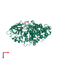
homo tetramer (preferred)
Assembly name:
Putative acyl-CoA dehydrogenase AidB (preferred)
PDBe Complex ID:
PDB-CPX-152563 (preferred)
Entry contents:
1 distinct polypeptide molecule
Macromolecule:


