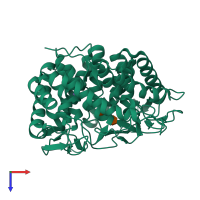
4ayp

X-ray diffraction
0.85Å resolution

Structure of The GH47 processing alpha-1,2-mannosidase from Caulobacter strain K31 in complex with thiomannobioside

Released:

Function and Biology Details

Reaction catalysed:
(1a) Man(9)GlcNAc(2)-[protein] + H(2)O = Man(8)GlcNAc(2)-[protein] (isomer 8A(1,2,3)B(1,2)) + beta-D-mannopyranose
Biochemical function:
Biological process:
Cellular component:

Structure analysis Details

Assembly composition:
monomeric (preferred)
PDBe Complex ID:
PDB-CPX-109212 (preferred)
Entry contents:
1 distinct polypeptide molecule
Macromolecules (2 distinct):
Mannosyl-oligosaccharide 1,2-alpha-mannosidase Chain: A
Molecule details ›
Chain: A
Length: 447 amino acids
Theoretical weight: 50.55 KDa
Source organism: Caulobacter sp. K31
Expression system: Escherichia coli BL21(DE3)
UniProt:
  • Canonical: B0SWV2 (Residues: 27-462; Coverage: 100%)
Gene name: Caul_4035
Sequence domains: Glycosyl hydrolase family 47
Structure domains: Glycosyltransferase

Ligands and Environments

Carbohydrate polymer : NEW Components: Z5L, MAN
2 bound ligands:
No modified residues

Experiments and Validation Details

Entry percentile scores
X-ray source: DIAMOND BEAMLINE I03
Spacegroup: R3
Unit cell:
a: 144.135Å b: 144.135Å c: 50.301Å
α: 90° β: 90° γ: 120°
R-values:
R R work R free
0.097 0.096 0.106
Expression system: Escherichia coli BL21(DE3)