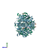
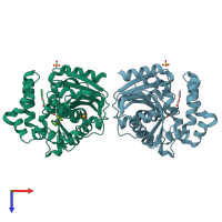
4o08

X-ray diffraction
1.95Å resolution

Crystal structure of bacillus megaterium epoxide hydrolase in complex with an inhibitor

Released:
Source organism: Priestia megaterium
Primary publication:
Engineering of an epoxide hydrolase for efficient bioresolution of bulky pharmaco substrates.
Proc Natl Acad Sci U S A 111 15717-22 (2014)
PMID: 25331869

Function and Biology Details

Reaction catalysed:
A pyrimidine 5'-nucleotide + H(2)O = D-ribose 5-phosphate + a pyrimidine base
Biological process:
  • not assigned
Cellular component:
  • not assigned

Structure analysis Details

Assembly composition:
monomeric (preferred)
PDBe Complex ID:
PDB-CPX-124658 (preferred)
Entry contents:
1 distinct polypeptide molecule
Macromolecule:
AB hydrolase-1 domain-containing protein Chains: A, B
Molecule details ›
Chains: A, B
Length: 321 amino acids
Theoretical weight: 37.18 KDa
Source organism: Priestia megaterium
Expression system: Escherichia coli BL21
UniProt:
  • Canonical: G9BEX6 (Residues: 1-287; Coverage: 100%)
Sequence domains: alpha/beta hydrolase fold
Structure domains: alpha/beta hydrolase

Ligands and Environments

2 bound ligands:
No modified residues

Experiments and Validation Details

Entry percentile scores
X-ray source: RIGAKU MICROMAX-007 HF
Spacegroup: P41212
Unit cell:
a: 108.541Å b: 108.541Å c: 116.843Å
α: 90° β: 90° γ: 90°
R-values:
R R work R free
0.171 0.17 0.207
Expression system: Escherichia coli BL21