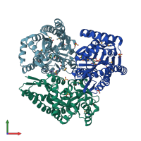
4pbq

X-ray diffraction
1.65Å resolution

CRYSTAL STRUCTURE OF A TRAP PERIPLASMIC SOLUTE BINDING PROTEIN FROM HAEMOPHILUS INFLUENZAE RdAW (HICG_00826, TARGET EFI-510123) WITH BOUND L-GULONATE

Released:
Model geometry
Fit model/data

Function and Biology Details

Structure analysis Details

Assembly composition:
monomeric (preferred)
PDBe Complex ID:
PDB-CPX-165007 (preferred)
Entry contents:
1 distinct polypeptide molecule
Macromolecule:
Putative TRAP PERIPLASMIC SOLUTE BINDING PROTEIN Chains: A, B, C
Molecule details ›
Chains: A, B, C
Length: 327 amino acids
Theoretical weight: 38.28 KDa
Source organism: Haemophilus influenzae RdAW
Expression system: Escherichia coli BL21(DE3)
Structure domains: Bacterial extracellular solute-binding protein, family 7

Ligands and Environments

1 modified residue:

Experiments and Validation Details

wwPDB Validation report is not available for this entry.
X-ray source: APS BEAMLINE 31-ID
Spacegroup: C2
Unit cell:
a: 133.449Å b: 76.537Å c: 112.977Å
α: 90° β: 114.01° γ: 90°
R-values:
R R work R free
0.149 0.147 0.184
Expression system: Escherichia coli BL21(DE3)