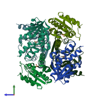
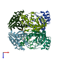
4r0n

X-ray diffraction
2Å resolution

Hexagonal form of phosphopantetheine adenylyltransferase from Mycobacterium tuberculosis

Released:
Entry authors: Timofeev VI, Smirnova EA, Chupova LA, Esipov RS, Kuranova IP

Function and Biology Details

Structure analysis Details

Assembly composition:
homo hexamer (preferred)
PDBe Complex ID:
PDB-CPX-165131 (preferred)
Entry contents:
1 distinct polypeptide molecule
Macromolecule:
Phosphopantetheine adenylyltransferase Chains: A, C, E, G, I, K
Molecule details ›
Chains: A, C, E, G, I, K
Length: 156 amino acids
Theoretical weight: 17.03 KDa
Source organism: Mycobacterium tuberculosis BT1
Expression system: Escherichia coli
Structure domains: HUPs

Ligands and Environments

1 bound ligand:
No modified residues

Experiments and Validation Details

Entry percentile scores
X-ray source: SPRING-8 BEAMLINE BL41XU
Spacegroup: P32
Unit cell:
a: 106.472Å b: 106.472Å c: 71.323Å
α: 90° β: 90° γ: 120°
R-values:
R R work R free
0.213 0.21 0.266
Expression system: Escherichia coli