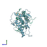
5pzi

X-ray diffraction
1.62Å resolution

PanDDA analysis group deposition -- Crystal Structure of SP100 after initial refinement with no ligand modelled (structure 114)

Released:

Function and Biology Details

Structure analysis Details

Assembly composition:
monomeric (preferred)
PDBe Complex ID:
PDB-CPX-540502 (preferred)
Entry contents:
1 distinct polypeptide molecule
Macromolecule:
Nuclear autoantigen Sp-100 Chains: A, B
Molecule details ›
Chains: A, B
Length: 180 amino acids
Theoretical weight: 21.5 KDa
Source organism: Homo sapiens
Expression system: Escherichia coli
UniProt:
  • Canonical: P23497 (Residues: 466-498, 499-508; Coverage: 5%)
  • Best match: P23497-4 (Residues: 700-879)
Gene name: SP100
Structure domains:

Ligands and Environments

3 bound ligands:
No modified residues

Experiments and Validation Details

Entry percentile scores
X-ray source: DIAMOND BEAMLINE I04-1
Spacegroup: C2
Unit cell:
a: 127.751Å b: 45.441Å c: 83.437Å
α: 90° β: 102.22° γ: 90°
R-values:
R R work R free
0.173 0.172 0.203
Expression system: Escherichia coli