

-L-glutamic acid</span>;</li> <li class='image_legend_li'>2 copies of <span class='highlight'>PYRIMIDINE-2,4-DIAMINE</span>;</li> <li class='image_legend_li'>2 copies of <span class='highlight'>NADP NICOTINAMIDE-ADENINE-DINUCLEOTIDE PHOSPHATE</span>;</li> <li class='image_legend_li'>2 copies of <span class='highlight'>SODIUM ION</span>;</li> <li class='image_legend_li'>1 copy of <span class='highlight'>water</span>.</li></ul>")
-L-glutamic acid</span>;</li> <li class='image_legend_li'>2 copies of <span class='highlight'>PYRIMIDINE-2,4-DIAMINE</span>;</li> <li class='image_legend_li'>2 copies of <span class='highlight'>NADP NICOTINAMIDE-ADENINE-DINUCLEOTIDE PHOSPHATE</span>;</li> <li class='image_legend_li'>2 copies of <span class='highlight'>SODIUM ION</span>;</li> <li class='image_legend_li'>1 copy of <span class='highlight'>water</span>.</li></ul>")
-L-glutamic acid</span>;</li> <li class='image_legend_li'>2 copies of <span class='highlight'>PYRIMIDINE-2,4-DIAMINE</span>;</li> <li class='image_legend_li'>2 copies of <span class='highlight'>NADP NICOTINAMIDE-ADENINE-DINUCLEOTIDE PHOSPHATE</span>;</li> <li class='image_legend_li'>2 copies of <span class='highlight'>SODIUM ION</span>;</li> <li class='image_legend_li'>1 copy of <span class='highlight'>water</span>.</li></ul>")
Function and Biology Details
Reaction catalysed:
5,6,7,8-tetrahydrofolate + NADP(+) = 7,8-dihydrofolate + NADPH
Biochemical function:
Biological process:
Cellular component:
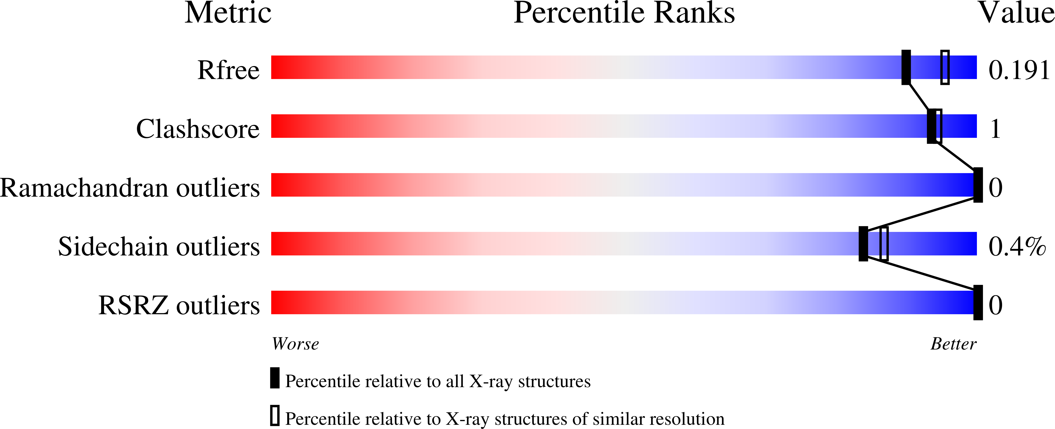
Structure analysis Details
Assembly composition:
monomeric (preferred)
Assembly name:
Dihydrofolate reductase (preferred)
PDBe Complex ID:
PDB-CPX-142189 (preferred)
Entry contents:
1 distinct polypeptide molecule
Macromolecule:
Dihydrofolate reductase
Molecule details ›
Chains: A, B
Length: 163 amino acids
Theoretical weight: 18.16 KDa
Source organism: Escherichia coli
Expression system: Escherichia coli
UniProt:
Sequence domains: Dihydrofolate reductase
Structure domains: Dihydrofolate Reductase, subunit A
Length: 163 amino acids
Theoretical weight: 18.16 KDa
Source organism: Escherichia coli
Expression system: Escherichia coli
UniProt:
- Canonical:
 P0ABQ4 (Residues: 2-159; Coverage: 99%)
Sequence domains: Dihydrofolate reductase
Structure domains: Dihydrofolate Reductase, subunit A


