

-TARTARIC ACID</span>;</li> <li class='image_legend_li'>1 copy of <span class='highlight'>water</span>.</li></ul>")
-TARTARIC ACID</span>;</li> <li class='image_legend_li'>1 copy of <span class='highlight'>water</span>.</li></ul>")
-TARTARIC ACID</span>;</li> <li class='image_legend_li'>1 copy of <span class='highlight'>water</span>.</li></ul>")
Function and Biology Details
Reaction catalysed:
UDP-alpha-D-glucose + D-glucose 6-phosphate = UDP + alpha,alpha-trehalose 6-phosphate
Biochemical function:
Biological process:
Cellular component:
- not assigned
Sequence domains:
Structure analysis Details
Assembly composition:
monomeric (preferred)
Assembly name:
Trehalose-6-phosphate synthase (preferred)
PDBe Complex ID:
PDB-CPX-171887 (preferred)
Entry contents:
1 distinct polypeptide molecule
Macromolecule:
Trehalose-6-phosphate synthase
Molecule details ›
Chains: A, B, C, D, E, F, G, H
Length: 494 amino acids
Theoretical weight: 55.18 KDa
Source organism: Paraburkholderia xenovorans LB400
Expression system: Escherichia coli BL21(DE3)
UniProt:
Sequence domains: Glycosyltransferase family 20
Structure domains: Glycogen Phosphorylase B;
Length: 494 amino acids
Theoretical weight: 55.18 KDa
Source organism: Paraburkholderia xenovorans LB400
Expression system: Escherichia coli BL21(DE3)
UniProt:
- Canonical:
 Q13W28 (Residues: 1-486; Coverage: 100%)
Sequence domains: Glycosyltransferase family 20
Structure domains: Glycogen Phosphorylase B;
Ligands and Environments


Experiments and Validation Details
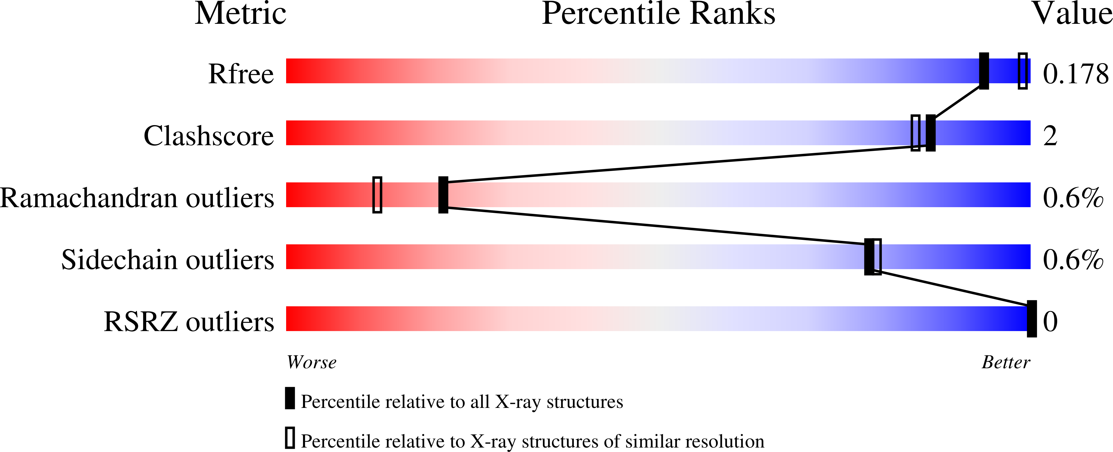
wwPDB Validation report is not available for this entry.
X-ray source:
APS BEAMLINE 21-ID-F
Spacegroup:
P1
Expression system: Escherichia coli BL21(DE3)
