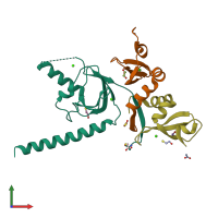
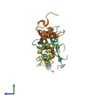
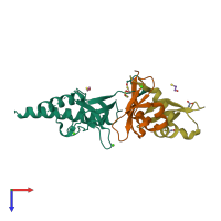
6hw2

X-ray diffraction
1.94Å resolution

The Crystal Structure of CaV beta4c in complex with HP1gamma chromo shadow domains

Released:
Source organisms:
Entry authors: Tanner N, Tripathy DR, Hirsch JA

Function and Biology Details

Structure analysis Details

Assembly composition:
hetero trimer (preferred)
PDBe Complex ID:
PDB-CPX-112610 (preferred)
Entry contents:
2 distinct polypeptide molecules
Macromolecules (2 distinct):
Voltage-dependent L-type calcium channel subunit beta-4 Chain: A
Molecule details ›
Chain: A
Length: 154 amino acids
Theoretical weight: 17.53 KDa
Source organism: Rattus norvegicus
Expression system: Escherichia coli 'BL21-Gold(DE3)pLysS AG'
UniProt:
  • Canonical: D4A055 (Residues: 56-232; Coverage: 30%)
Gene name: Cacnb4
Sequence domains: Voltage gated calcium channel subunit beta domain 4Aa N terminal
Chromobox protein homolog 3 Chains: B, C
Molecule details ›
Chains: B, C
Length: 75 amino acids
Theoretical weight: 8.47 KDa
Source organism: Homo sapiens
Expression system: Escherichia coli 'BL21-Gold(DE3)pLysS AG'
UniProt:
  • Canonical: Q13185 (Residues: 109-183; Coverage: 41%)
Gene name: CBX3
Sequence domains: Chromo shadow domain

Ligands and Environments

No modified residues

Experiments and Validation Details

Entry percentile scores
X-ray source: ESRF BEAMLINE ID14-2
Spacegroup: C2
Unit cell:
a: 115.026Å b: 58.036Å c: 57.546Å
α: 90° β: 109.7° γ: 90°
R-values:
R R work R free
0.179 0.178 0.21
Expression system: Escherichia coli 'BL21-Gold(DE3)pLysS AG'