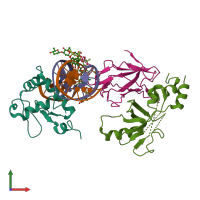
6vgg

X-ray diffraction
4.31Å resolution

Crystal structure of the DNA binding domains of human transcription factor ERG, human Runx2 bound to core binding factor beta (Cbfb), and mithramycin, in complex with 16mer DNA CAGAGGATGTGGCTTC

Released:
Model geometry
Data not analysed
Fit model/data
Data not analysed
Source organism: Homo sapiens
Primary publication:
Allosteric interference in oncogenic FLI1 and ERG transactions by mithramycins.
Structure 29 404-412.e4 (2021)
PMID: 33275876

Function and Biology Details

Structure analysis Details

Assembly composition:
hetero pentamer (preferred)
PDBe Complex ID:
PDB-CPX-120775 (preferred)
Entry contents:
3 distinct polypeptide molecules
2 distinct DNA molecules
Macromolecules (5 distinct):
Transcriptional regulator ERG Chain: A
Molecule details ›
Chain: A
Length: 128 amino acids
Theoretical weight: 14.93 KDa
Source organism: Homo sapiens
Expression system: Escherichia coli BL21(DE3)
UniProt:
  • Canonical: P11308 (Residues: 306-419; Coverage: 24%)
Gene name: ERG
Sequence domains: Ets-domain
Runt-related transcription factor 2 Chain: D
Molecule details ›
Chain: D
Length: 177 amino acids
Theoretical weight: 19.54 KDa
Source organism: Homo sapiens
Expression system: Escherichia coli BL21(DE3)
UniProt:
  • Canonical: Q13950 (Residues: 111-287; Coverage: 34%)
Gene names: AML3, CBFA1, OSF2, PEBP2A, RUNX2
Sequence domains: Runt domain
Core-binding factor subunit beta Chain: G
DNA (5'-D(P*CP*AP*GP*AP*GP*GP*AP*TP*GP*TP*GP*GP*CP*TP*TP*C)-3') Chain: B
DNA (5'-D(P*GP*AP*AP*GP*CP*CP*AP*CP*AP*TP*CP*CP*TP*CP*TP*G)-3') Chain: C

Ligands and Environments

2 bound ligands:
No modified residues

Experiments and Validation Details

wwPDB Validation report is not available for this entry.
X-ray source: APS BEAMLINE 22-ID
Spacegroup: P6222
Unit cell:
a: 104.678Å b: 104.678Å c: 322.836Å
α: 90° β: 90° γ: 120°
R-values:
R R work R free
0.253 0.251 0.285
Expression system: Escherichia coli BL21(DE3)