EMD-10388
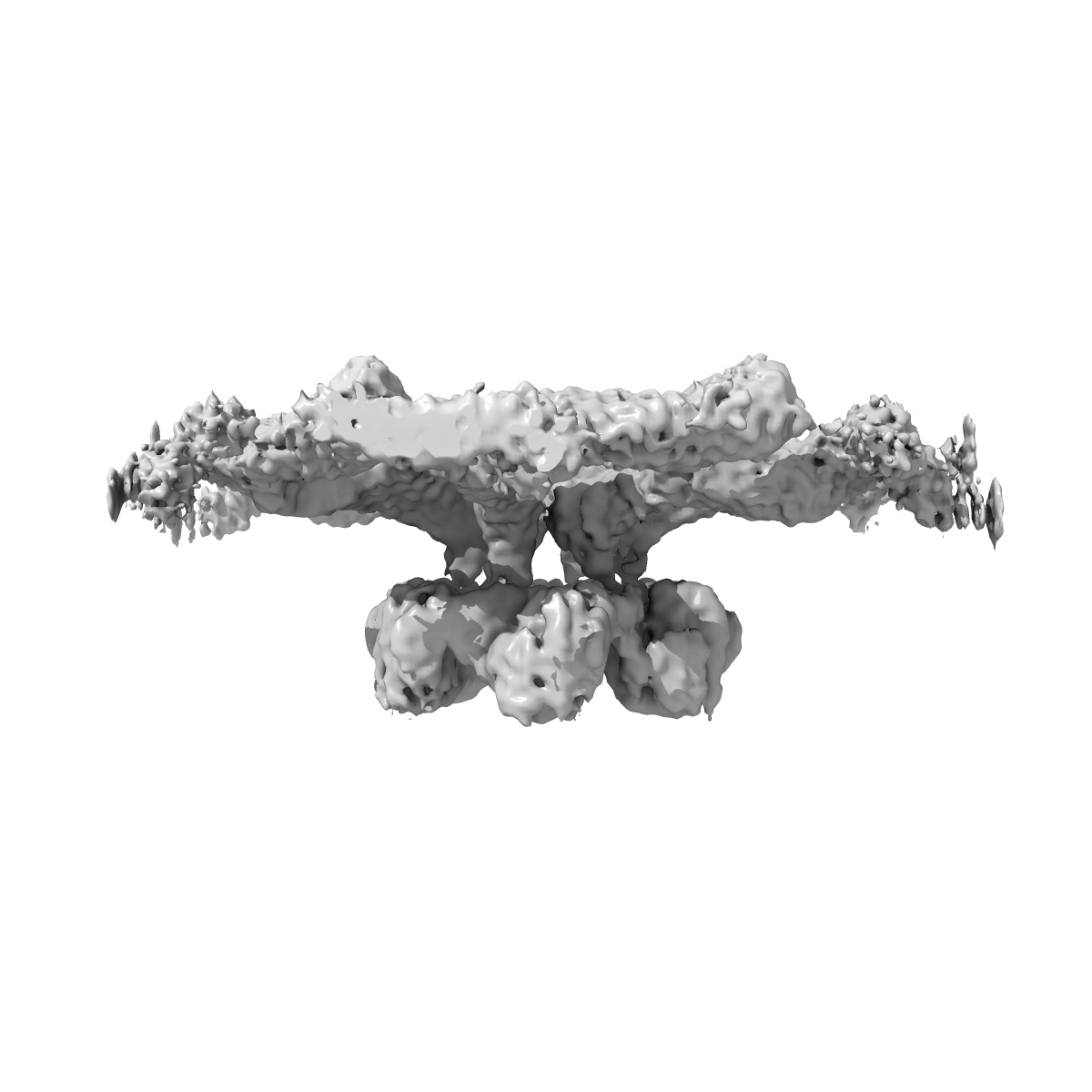
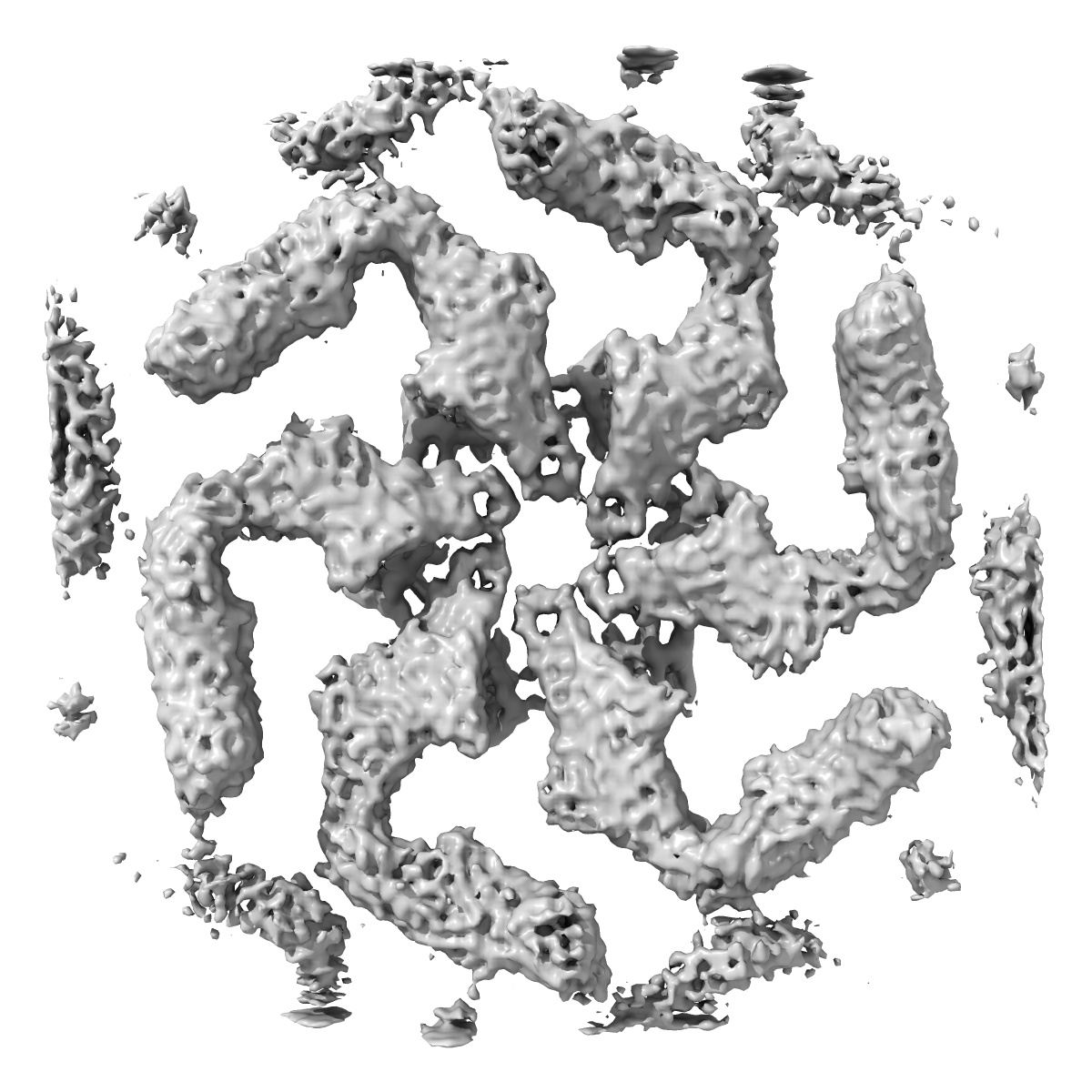
In situ structure of the Caulobacter crescentus S-layer
EMD-10388
Subtomogram averaging4.8 Å
 Deposition: 21/10/2019
Map released: 15/01/2020
Last modified: 29/07/2020
Sample Organism:
Caulobacter crescentus NA1000
Sample: Caulobacter crescentus S-layer
Deposition Authors: Bharat T, von Kuegelgen A
Sample: Caulobacter crescentus S-layer
Deposition Authors: Bharat T, von Kuegelgen A
In Situ Structure of an Intact Lipopolysaccharide-Bound Bacterial Surface Layer.
von Kugelgen A  ,
Tang H ,
Hardy GG ,
Kureisaite-Ciziene D,
Brun YV,
Stansfeld PJ ,
Robinson CV,
Bharat TAM
,
Tang H ,
Hardy GG ,
Kureisaite-Ciziene D,
Brun YV,
Stansfeld PJ ,
Robinson CV,
Bharat TAM
(2020) Cell , 180 , 348 - 358.e15
 ,
Tang H ,
Hardy GG ,
Kureisaite-Ciziene D,
Brun YV,
Stansfeld PJ ,
Robinson CV,
Bharat TAM
,
Tang H ,
Hardy GG ,
Kureisaite-Ciziene D,
Brun YV,
Stansfeld PJ ,
Robinson CV,
Bharat TAM
(2020) Cell , 180 , 348 - 358.e15
Abstract:
Most bacterial and all archaeal cells are encapsulated by a paracrystalline, protective, and cell-shape-determining proteinaceous surface layer (S-layer). On Gram-negative bacteria, S-layers are anchored to cells via lipopolysaccharide. Here, we report an electron cryomicroscopy structure of the Caulobacter crescentus S-layer bound to the O-antigen of lipopolysaccharide. Using native mass spectrometry and molecular dynamics simulations, we deduce the length of the O-antigen on cells and show how lipopolysaccharide binding and S-layer assembly is regulated by calcium. Finally, we present a near-atomic resolution in situ structure of the complete S-layer using cellular electron cryotomography, showing S-layer arrangement at the tip of the O-antigen. A complete atomic structure of the S-layer shows the power of cellular tomography for in situ structural biology and sheds light on a very abundant class of self-assembling molecules with important roles in prokaryotic physiology with marked potential for synthetic biology and surface-display applications.
Most bacterial and all archaeal cells are encapsulated by a paracrystalline, protective, and cell-shape-determining proteinaceous surface layer (S-layer). On Gram-negative bacteria, S-layers are anchored to cells via lipopolysaccharide. Here, we report an electron cryomicroscopy structure of the Caulobacter crescentus S-layer bound to the O-antigen of lipopolysaccharide. Using native mass spectrometry and molecular dynamics simulations, we deduce the length of the O-antigen on cells and show how lipopolysaccharide binding and S-layer assembly is regulated by calcium. Finally, we present a near-atomic resolution in situ structure of the complete S-layer using cellular electron cryotomography, showing S-layer arrangement at the tip of the O-antigen. A complete atomic structure of the S-layer shows the power of cellular tomography for in situ structural biology and sheds light on a very abundant class of self-assembling molecules with important roles in prokaryotic physiology with marked potential for synthetic biology and surface-display applications.