EMD-10464
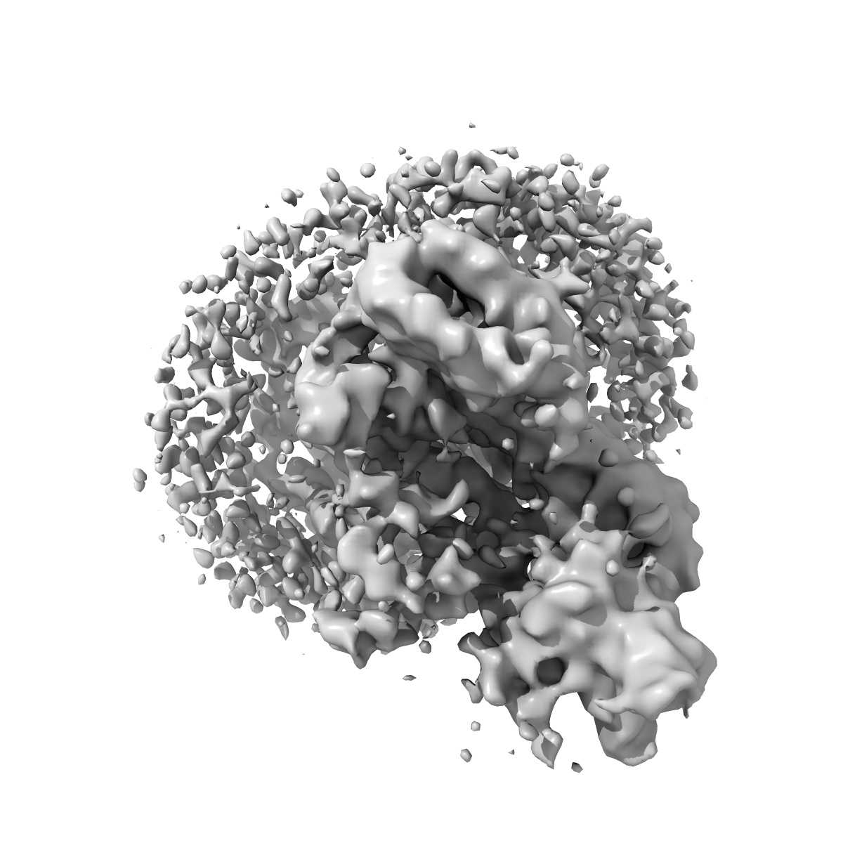
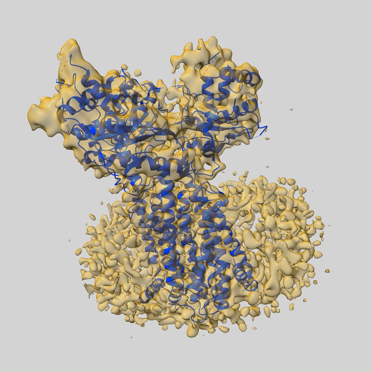
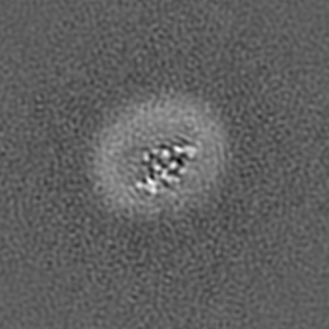
Structure of Drosophila melanogaster Dispatched bound to a modified Hedgehog ligand, HhN-C85II
EMD-10464
Single-particle4.76 Å
 Deposition: 07/11/2019
Map released: 03/06/2020
Last modified: 23/10/2024
Sample Organism:
Drosophila melanogaster
Sample: A complex of Dispatched and HhN-C85II in digitonin micelle
Fitted models: 6td6 (Avg. Q-score: 0.263)
Deposition Authors: Korkhov VM ,
Cannac F
,
Cannac F
Sample: A complex of Dispatched and HhN-C85II in digitonin micelle
Fitted models: 6td6 (Avg. Q-score: 0.263)
Deposition Authors: Korkhov VM
 ,
Cannac F
,
Cannac F
Cryo-EM structure of the Hedgehog release protein Dispatched.
Cannac F ,
Qi C ,
Falschlunger J ,
Hausmann G,
Basler K ,
Korkhov VM
(2020) Sci Adv , 6 , eaay7928 - eaay7928
,
Qi C ,
Falschlunger J ,
Hausmann G,
Basler K ,
Korkhov VM
(2020) Sci Adv , 6 , eaay7928 - eaay7928
Abstract:
The Hedgehog (Hh) signaling pathway controls embryonic development and adult tissue homeostasis in multicellular organisms. In Drosophila melanogaster, the pathway is primed by secretion of a dually lipid-modified morphogen, Hh, a process dependent on a membrane-integral protein Dispatched. Although Dispatched is a critical component of the pathway, the structural basis of its activity has, so far, not been described. Here, we describe a cryo-electron microscopy structure of the D. melanogaster Dispatched at 3.2-Å resolution. The ectodomains of Dispatched adopt an open conformation suggestive of a receptor-chaperone role. A three-dimensional reconstruction of Dispatched bound to Hh confirms the ability of Dispatched to bind Hh but using a unique mode distinct from those previously observed in structures of Hh complexes. The structure may represent the state of the complex that precedes shedding of Hh from the surface of the morphogen-releasing cell.
The Hedgehog (Hh) signaling pathway controls embryonic development and adult tissue homeostasis in multicellular organisms. In Drosophila melanogaster, the pathway is primed by secretion of a dually lipid-modified morphogen, Hh, a process dependent on a membrane-integral protein Dispatched. Although Dispatched is a critical component of the pathway, the structural basis of its activity has, so far, not been described. Here, we describe a cryo-electron microscopy structure of the D. melanogaster Dispatched at 3.2-Å resolution. The ectodomains of Dispatched adopt an open conformation suggestive of a receptor-chaperone role. A three-dimensional reconstruction of Dispatched bound to Hh confirms the ability of Dispatched to bind Hh but using a unique mode distinct from those previously observed in structures of Hh complexes. The structure may represent the state of the complex that precedes shedding of Hh from the surface of the morphogen-releasing cell.