EMD-10467
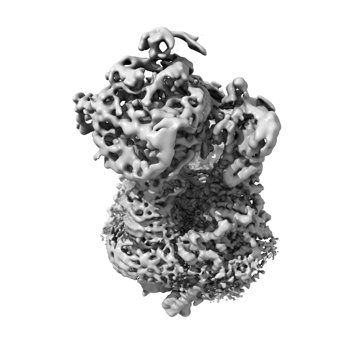
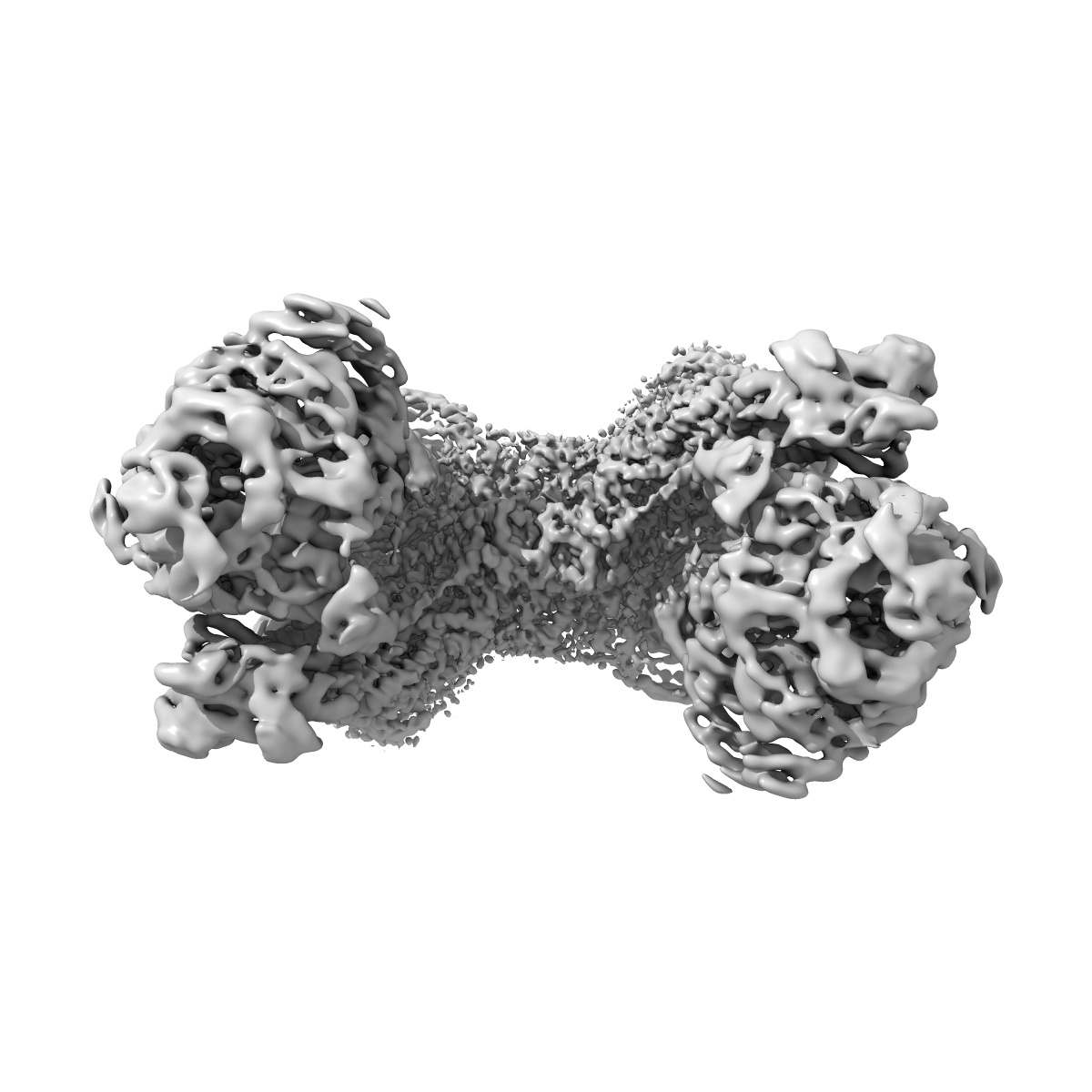
Cryo-EM structure of Euglena gracilis mitochondrial ATP synthase, full dimer, rotational states 1
EMD-10467
Single-particle4.32 Å
 Deposition: 10/11/2019
Map released: 27/11/2019
Last modified: 23/10/2024
Sample Organism:
Euglena gracilis
Sample: Euglena gracilis mitochondrial ATP synthase dimer
Fitted models: 6tdu (Avg. Q-score: 0.236)
Deposition Authors: Muhleip A ,
Amunts A
,
Amunts A
Sample: Euglena gracilis mitochondrial ATP synthase dimer
Fitted models: 6tdu (Avg. Q-score: 0.236)
Deposition Authors: Muhleip A
 ,
Amunts A
,
Amunts A
Structure of a mitochondrial ATP synthase with bound native cardiolipin.
Abstract:
The mitochondrial ATP synthase fuels eukaryotic cells with chemical energy. Here we report the cryo-EM structure of a divergent ATP synthase dimer from mitochondria of Euglena gracilis, a member of the phylum Euglenozoa that also includes human parasites. It features 29 different subunits, 8 of which are newly identified. The membrane region was determined to 2.8 Å resolution, enabling the identification of 37 associated lipids, including 25 cardiolipins, which provides insight into protein-lipid interactions and their functional roles. The rotor-stator interface comprises four membrane-embedded horizontal helices, including a distinct subunit a. The dimer interface is formed entirely by phylum-specific components, and a peripherally associated subcomplex contributes to the membrane curvature. The central and peripheral stalks directly interact with each other. Last, the ATPase inhibitory factor 1 (IF1) binds in a mode that is different from human, but conserved in Trypanosomatids.
The mitochondrial ATP synthase fuels eukaryotic cells with chemical energy. Here we report the cryo-EM structure of a divergent ATP synthase dimer from mitochondria of Euglena gracilis, a member of the phylum Euglenozoa that also includes human parasites. It features 29 different subunits, 8 of which are newly identified. The membrane region was determined to 2.8 Å resolution, enabling the identification of 37 associated lipids, including 25 cardiolipins, which provides insight into protein-lipid interactions and their functional roles. The rotor-stator interface comprises four membrane-embedded horizontal helices, including a distinct subunit a. The dimer interface is formed entirely by phylum-specific components, and a peripherally associated subcomplex contributes to the membrane curvature. The central and peripheral stalks directly interact with each other. Last, the ATPase inhibitory factor 1 (IF1) binds in a mode that is different from human, but conserved in Trypanosomatids.