EMD-11497
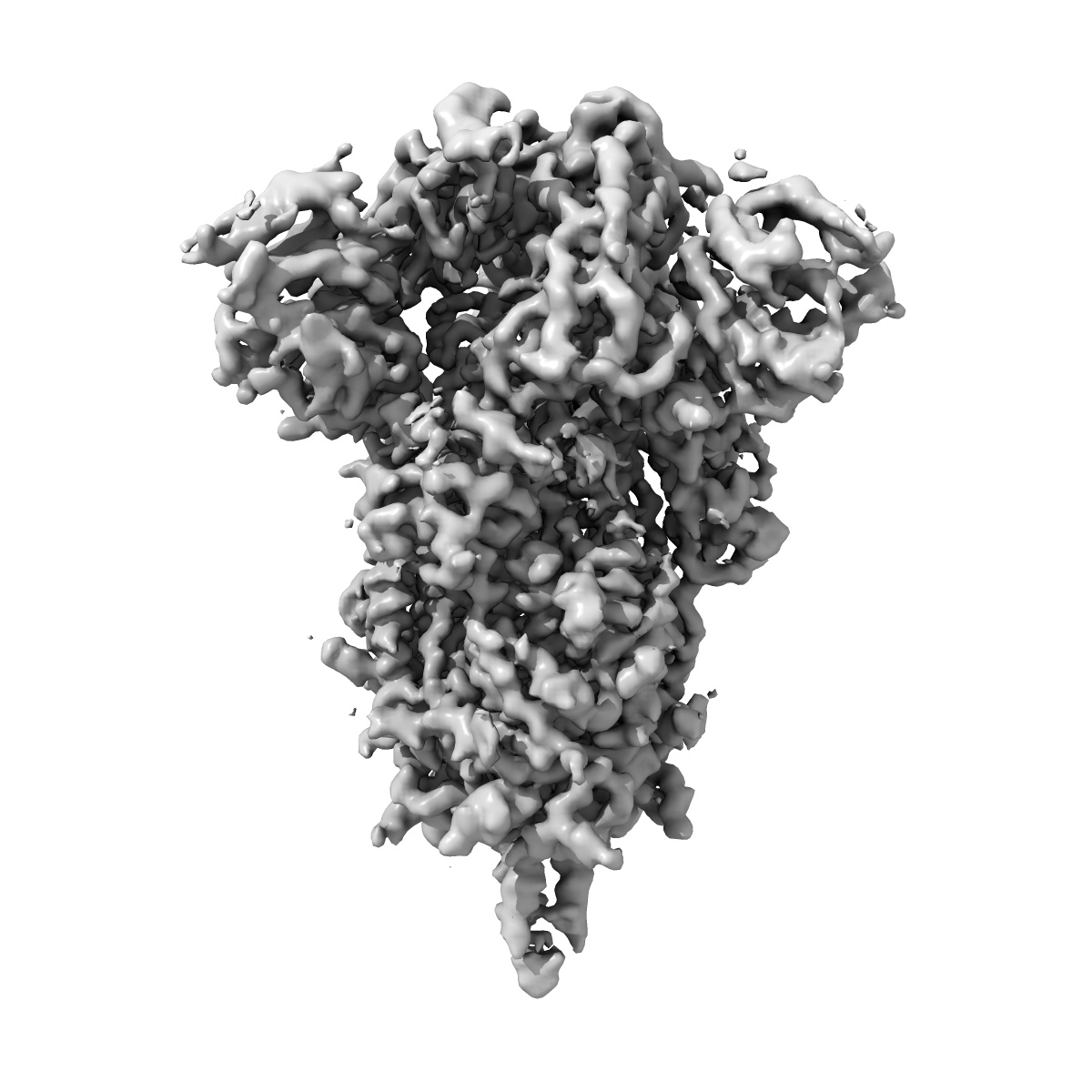
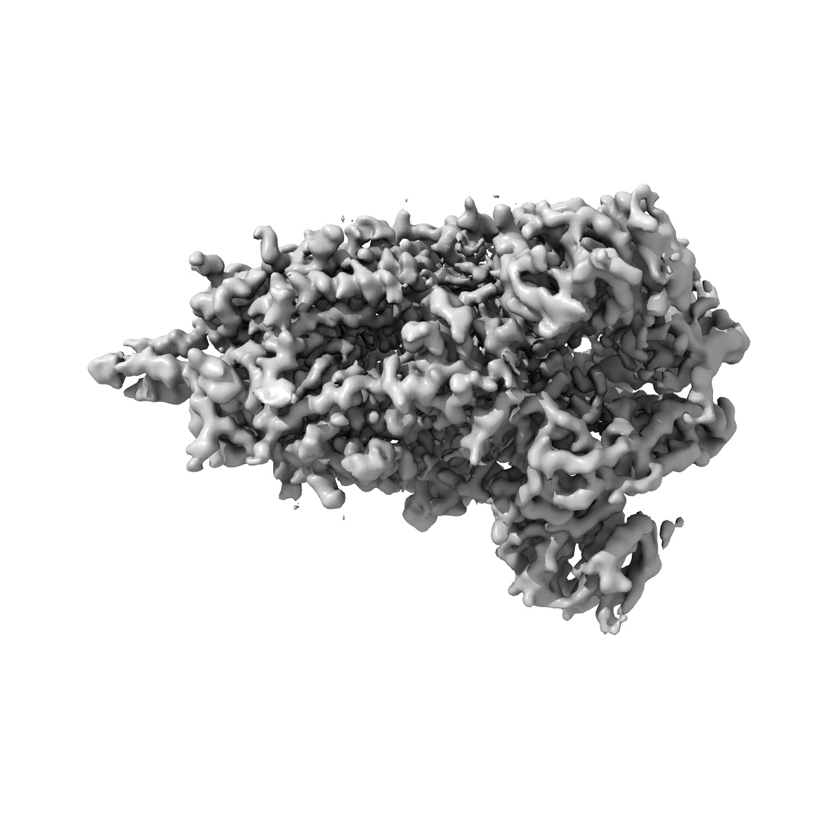
Cryo-EM structure of SARS-CoV-2 Spike Proteins on intact virions: 3 Closed RBDs
EMD-11497
Single-particle3.5 Å
 Deposition: 28/07/2020
Map released: 05/08/2020
Last modified: 23/10/2024
Sample Organism:
Severe acute respiratory syndrome coronavirus 2
Sample: Severe acute respiratory syndrome coronavirus 2
Fitted models: 6zwv (Avg. Q-score: 0.402)
Raw data: EMPIAR-10492
Deposition Authors: Ke Z ,
Qu K ,
Nakane T ,
Xiong X ,
Cortese M ,
Zila V ,
Scheres SHW ,
Briggs JAG
,
Qu K ,
Nakane T ,
Xiong X ,
Cortese M ,
Zila V ,
Scheres SHW ,
Briggs JAG
Sample: Severe acute respiratory syndrome coronavirus 2
Fitted models: 6zwv (Avg. Q-score: 0.402)
Raw data: EMPIAR-10492
Deposition Authors: Ke Z
 ,
Qu K ,
Nakane T ,
Xiong X ,
Cortese M ,
Zila V ,
Scheres SHW ,
Briggs JAG
,
Qu K ,
Nakane T ,
Xiong X ,
Cortese M ,
Zila V ,
Scheres SHW ,
Briggs JAG
Structures and distributions of SARS-CoV-2 spike proteins on intact virions.
Ke Z ,
Oton J ,
Qu K ,
Cortese M ,
Zila V ,
McKeane L,
Nakane T ,
Zivanov J,
Neufeldt CJ ,
Cerikan B,
Lu JM ,
Peukes J ,
Xiong X ,
Krausslich HG,
Scheres SHW ,
Bartenschlager R ,
Briggs JAG
(2020) Nature , 588 , 498 - 502
,
Oton J ,
Qu K ,
Cortese M ,
Zila V ,
McKeane L,
Nakane T ,
Zivanov J,
Neufeldt CJ ,
Cerikan B,
Lu JM ,
Peukes J ,
Xiong X ,
Krausslich HG,
Scheres SHW ,
Bartenschlager R ,
Briggs JAG
(2020) Nature , 588 , 498 - 502
Abstract:
Severe acute respiratory syndrome coronavirus 2 (SARS-CoV-2) virions are surrounded by a lipid bilayer from which spike (S) protein trimers protrude1. Heavily glycosylated S trimers bind to the angiotensin-converting enzyme 2 receptor and mediate entry of virions into target cells2-6. S exhibits extensive conformational flexibility: it modulates exposure of its receptor-binding site and subsequently undergoes complete structural rearrangement to drive fusion of viral and cellular membranes2,7,8. The structures and conformations of soluble, overexpressed, purified S proteins have been studied in detail using cryo-electron microscopy2,7,9-12, but the structure and distribution of S on the virion surface remain unknown. Here we applied cryo-electron microscopy and tomography to image intact SARS-CoV-2 virions and determine the high-resolution structure, conformational flexibility and distribution of S trimers in situ on the virion surface. These results reveal the conformations of S on the virion, and provide a basis from which to understand interactions between S and neutralizing antibodies during infection or vaccination.
Severe acute respiratory syndrome coronavirus 2 (SARS-CoV-2) virions are surrounded by a lipid bilayer from which spike (S) protein trimers protrude1. Heavily glycosylated S trimers bind to the angiotensin-converting enzyme 2 receptor and mediate entry of virions into target cells2-6. S exhibits extensive conformational flexibility: it modulates exposure of its receptor-binding site and subsequently undergoes complete structural rearrangement to drive fusion of viral and cellular membranes2,7,8. The structures and conformations of soluble, overexpressed, purified S proteins have been studied in detail using cryo-electron microscopy2,7,9-12, but the structure and distribution of S on the virion surface remain unknown. Here we applied cryo-electron microscopy and tomography to image intact SARS-CoV-2 virions and determine the high-resolution structure, conformational flexibility and distribution of S trimers in situ on the virion surface. These results reveal the conformations of S on the virion, and provide a basis from which to understand interactions between S and neutralizing antibodies during infection or vaccination.