EMD-14066
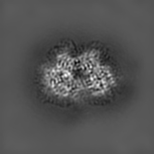
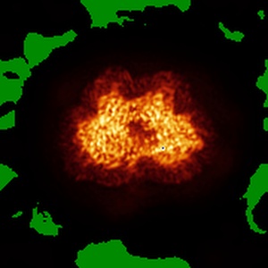
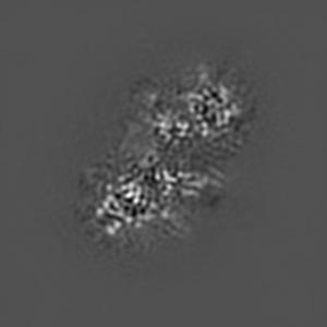
Structure of the Rab GEF complex Mon1-Ccz1
EMD-14066
Single-particle3.85 Å
 Deposition: 20/12/2021
Map released: 09/02/2022
Last modified: 17/07/2024
Sample Organism:
Chaetomium thermophilum
Sample: Rab GEF complex Mon1-Ccz1
Fitted models: 7qla (Avg. Q-score: 0.419)
Deposition Authors: Klink BU, Herrmann E
Sample: Rab GEF complex Mon1-Ccz1
Fitted models: 7qla (Avg. Q-score: 0.419)
Deposition Authors: Klink BU, Herrmann E

Structure of the Mon1-Ccz1 complex reveals molecular basis of membrane binding for Rab7 activation.
Klink BU,
Herrmann E ,
Antoni C,
Langemeyer L ,
Kiontke S ,
Gatsogiannis C ,
Ungermann C ,
Raunser S ,
Kummel D
(2022) PNAS , 119
,
Antoni C,
Langemeyer L ,
Kiontke S ,
Gatsogiannis C ,
Ungermann C ,
Raunser S ,
Kummel D
(2022) PNAS , 119
Abstract:
Activation of the GTPase Rab7/Ypt7 by its cognate guanine nucleotide exchange factor (GEF) Mon1-Ccz1 marks organelles such as endosomes and autophagosomes for fusion with lysosomes/vacuoles and degradation of their content. Here, we present a high-resolution cryogenic electron microscopy structure of the Mon1-Ccz1 complex that reveals its architecture in atomic detail. Mon1 and Ccz1 are arranged side by side in a pseudo-twofold symmetrical heterodimer. The three Longin domains of each Mon1 and Ccz1 are triangularly arranged, providing a strong scaffold for the catalytic center of the GEF. At the opposite side of the Ypt7-binding site, a positively charged and relatively flat patch stretches the Longin domains 2/3 of Mon1 and functions as a phosphatidylinositol phosphate-binding site, explaining how the GEF is targeted to membranes. Our work provides molecular insight into the mechanisms of endosomal Rab activation and serves as a blueprint for understanding the function of members of the Tri Longin domain Rab-GEF family.
Activation of the GTPase Rab7/Ypt7 by its cognate guanine nucleotide exchange factor (GEF) Mon1-Ccz1 marks organelles such as endosomes and autophagosomes for fusion with lysosomes/vacuoles and degradation of their content. Here, we present a high-resolution cryogenic electron microscopy structure of the Mon1-Ccz1 complex that reveals its architecture in atomic detail. Mon1 and Ccz1 are arranged side by side in a pseudo-twofold symmetrical heterodimer. The three Longin domains of each Mon1 and Ccz1 are triangularly arranged, providing a strong scaffold for the catalytic center of the GEF. At the opposite side of the Ypt7-binding site, a positively charged and relatively flat patch stretches the Longin domains 2/3 of Mon1 and functions as a phosphatidylinositol phosphate-binding site, explaining how the GEF is targeted to membranes. Our work provides molecular insight into the mechanisms of endosomal Rab activation and serves as a blueprint for understanding the function of members of the Tri Longin domain Rab-GEF family.