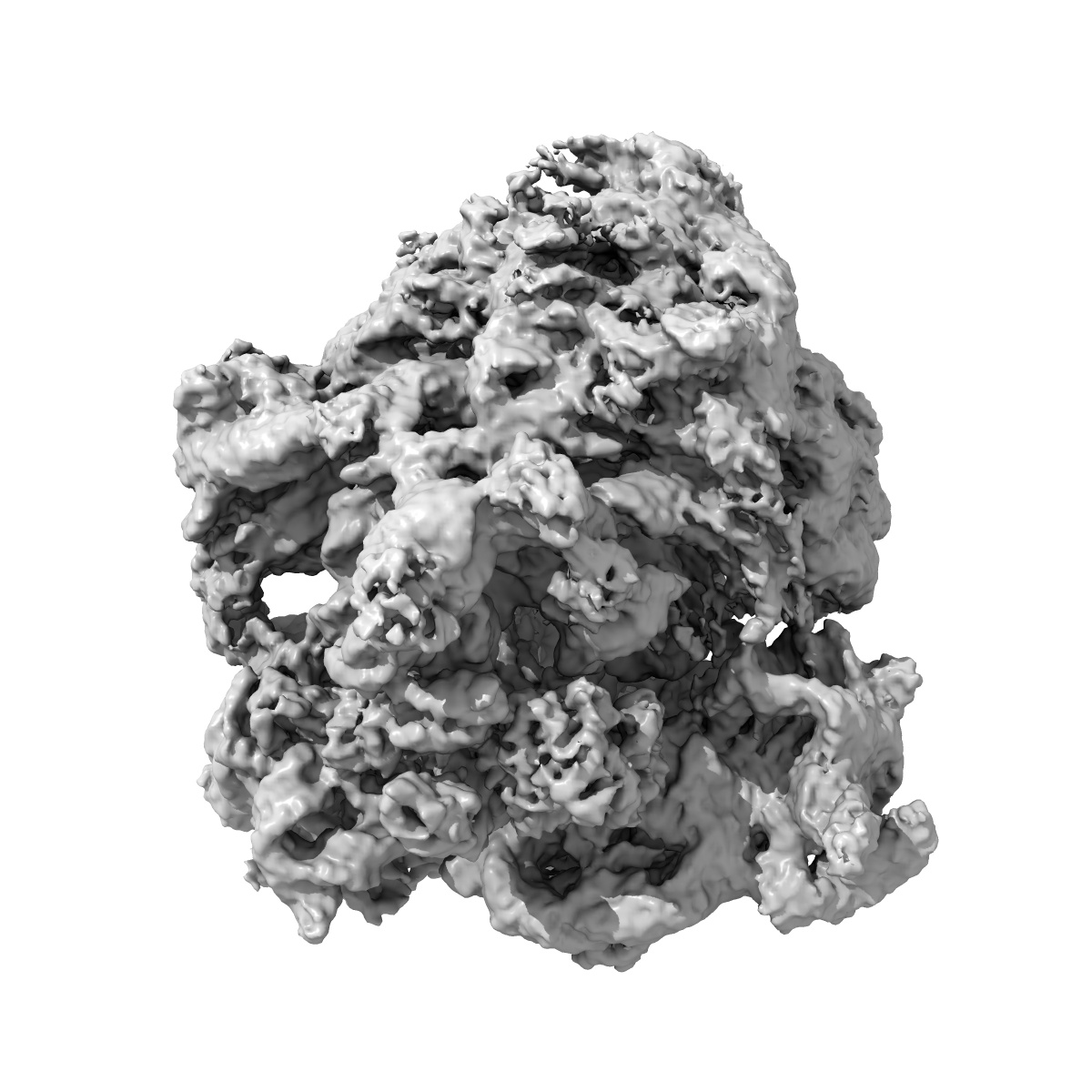
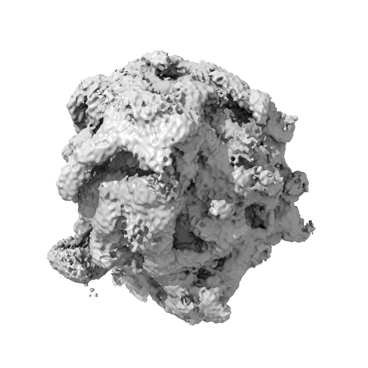
EMD-17664
Cami1-ribosome local refinement map with mask on Cami1 and L12-Cter
EMD-17664
Single-particle3.63 Å
 Deposition: 20/06/2023
Map released: 13/12/2023
Last modified: 27/03/2024
Sample Organism:
Escherichia coli
Sample: Cami1 bound in 70S E.coli ribosome
Deposition Authors: Tamulaitiene G ,
Mogila I ,
Sasnauskas G ,
Tamulaitis G
,
Mogila I ,
Sasnauskas G ,
Tamulaitis G
Sample: Cami1 bound in 70S E.coli ribosome
Deposition Authors: Tamulaitiene G
 ,
Mogila I ,
Sasnauskas G ,
Tamulaitis G
,
Mogila I ,
Sasnauskas G ,
Tamulaitis G
Ribosomal stalk-captured CARF-RelE ribonuclease inhibits translation following CRISPR signaling.
Mogila I ,
Tamulaitiene G ,
Keda K,
Timinskas A,
Ruksenaite A ,
Sasnauskas G ,
Venclovas C ,
Siksnys V ,
Tamulaitis G
(2023) Science , 382 , 1036 - 1041
,
Tamulaitiene G ,
Keda K,
Timinskas A,
Ruksenaite A ,
Sasnauskas G ,
Venclovas C ,
Siksnys V ,
Tamulaitis G
(2023) Science , 382 , 1036 - 1041
Abstract:
Prokaryotic type III CRISPR-Cas antiviral systems employ cyclic oligoadenylate (cAn) signaling to activate a diverse range of auxiliary proteins that reinforce the CRISPR-Cas defense. Here we characterize a class of cAn-dependent effector proteins named CRISPR-Cas-associated messenger RNA (mRNA) interferase 1 (Cami1) consisting of a CRISPR-associated Rossmann fold sensor domain fused to winged helix-turn-helix and a RelE-family mRNA interferase domain. Upon activation by cyclic tetra-adenylate (cA4), Cami1 cleaves mRNA exposed at the ribosomal A-site thereby depleting mRNA and leading to cell growth arrest. The structures of apo-Cami1 and the ribosome-bound Cami1-cA4 complex delineate the conformational changes that lead to Cami1 activation and the mechanism of Cami1 binding to a bacterial ribosome, revealing unexpected parallels with eukaryotic ribosome-inactivating proteins.
Prokaryotic type III CRISPR-Cas antiviral systems employ cyclic oligoadenylate (cAn) signaling to activate a diverse range of auxiliary proteins that reinforce the CRISPR-Cas defense. Here we characterize a class of cAn-dependent effector proteins named CRISPR-Cas-associated messenger RNA (mRNA) interferase 1 (Cami1) consisting of a CRISPR-associated Rossmann fold sensor domain fused to winged helix-turn-helix and a RelE-family mRNA interferase domain. Upon activation by cyclic tetra-adenylate (cA4), Cami1 cleaves mRNA exposed at the ribosomal A-site thereby depleting mRNA and leading to cell growth arrest. The structures of apo-Cami1 and the ribosome-bound Cami1-cA4 complex delineate the conformational changes that lead to Cami1 activation and the mechanism of Cami1 binding to a bacterial ribosome, revealing unexpected parallels with eukaryotic ribosome-inactivating proteins.