EMD-20284
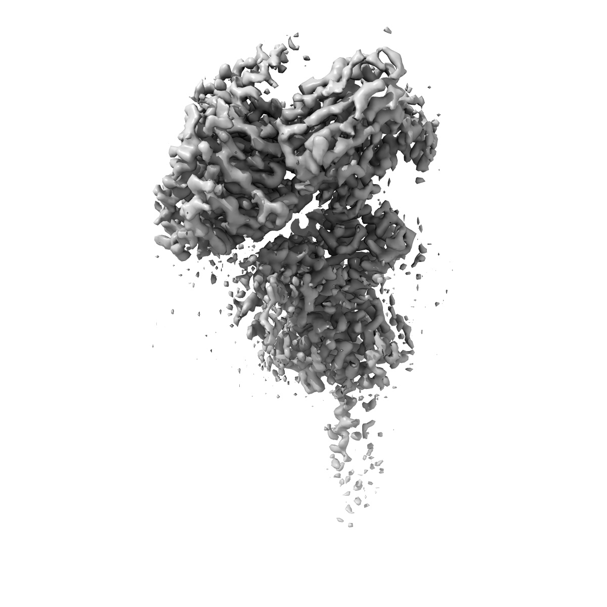
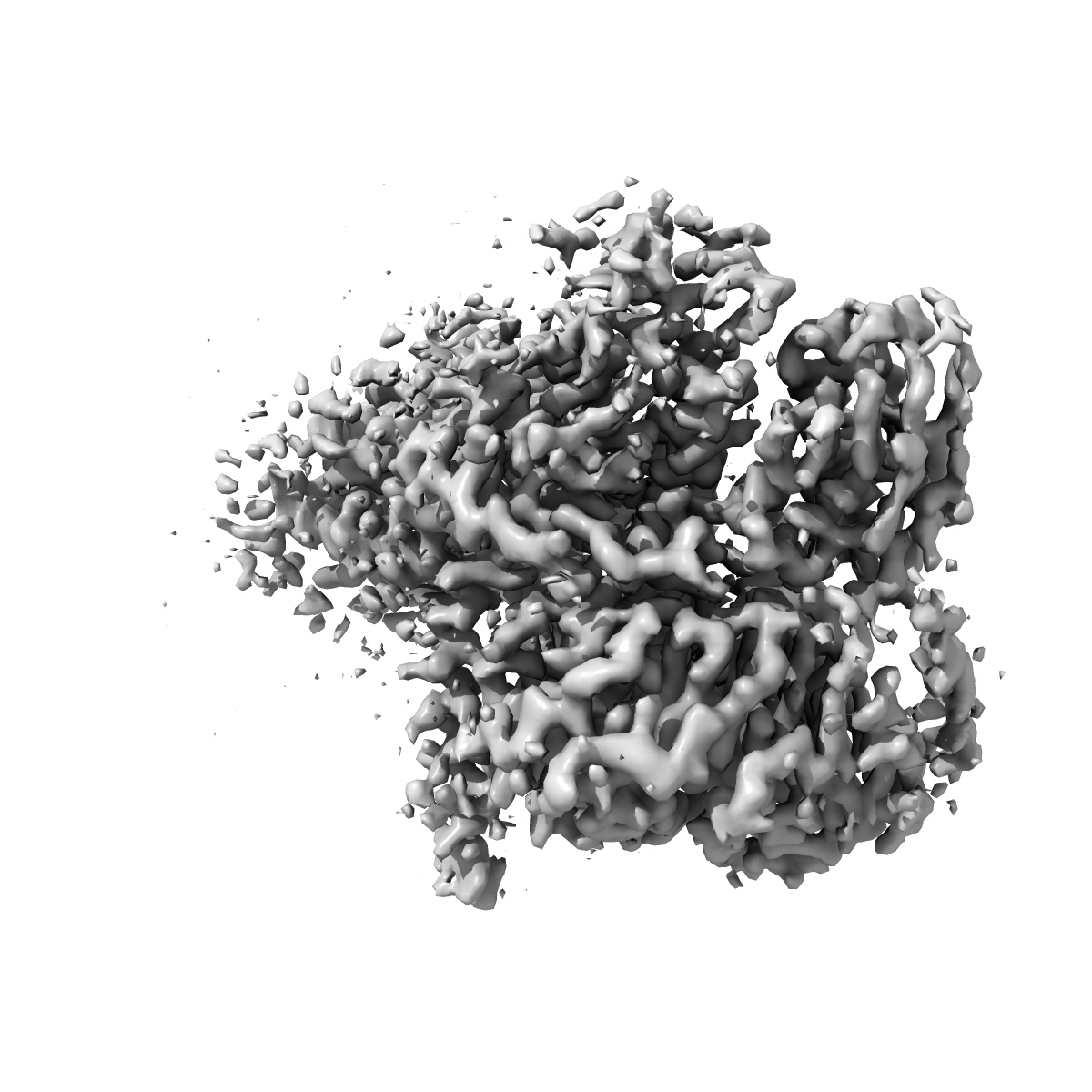
Cryo-EM structure of Urocortin 1-bound Corticotropin-releasing factor 1 receptor in complex with Gs protein and Nb35
EMD-20284
Single-particle3.0 Å
 Deposition: 12/06/2019
Map released: 12/02/2020
Last modified: 23/10/2024
Sample Organism:
Homo sapiens,
synthetic construct
Sample: Urocortin1-bound CRF1R in complex Gs and Nb35
Fitted models: 6pb0 (Avg. Q-score: 0.455)
Deposition Authors: Ma S, Shen Q
Sample: Urocortin1-bound CRF1R in complex Gs and Nb35
Fitted models: 6pb0 (Avg. Q-score: 0.455)
Deposition Authors: Ma S, Shen Q
Molecular Basis for Hormone Recognition and Activation of Corticotropin-Releasing Factor Receptors.
Ma S,
Shen Q,
Zhao LH,
Mao C  ,
Zhou XE,
Shen DD,
de Waal PW,
Bi P ,
Li C,
Jiang Y,
Wang MW,
Sexton PM,
Wootten D ,
Melcher K ,
Zhang Y ,
Xu HE
,
Zhou XE,
Shen DD,
de Waal PW,
Bi P ,
Li C,
Jiang Y,
Wang MW,
Sexton PM,
Wootten D ,
Melcher K ,
Zhang Y ,
Xu HE
(2020) Mol Cell , 77 , 669
 ,
Zhou XE,
Shen DD,
de Waal PW,
Bi P ,
Li C,
Jiang Y,
Wang MW,
Sexton PM,
Wootten D ,
Melcher K ,
Zhang Y ,
Xu HE
,
Zhou XE,
Shen DD,
de Waal PW,
Bi P ,
Li C,
Jiang Y,
Wang MW,
Sexton PM,
Wootten D ,
Melcher K ,
Zhang Y ,
Xu HE
(2020) Mol Cell , 77 , 669
Abstract:
Corticotropin-releasing factor (CRF) and the three related peptides urocortins 1-3 (UCN1-UCN3) are endocrine hormones that control the stress responses by activating CRF1R and CRF2R, two members of class B G-protein-coupled receptors (GPCRs). Here, we present two cryoelectron microscopy (cryo-EM) structures of UCN1-bound CRF1R and CRF2R with the stimulatory G protein. In both structures, UCN1 adopts a single straight helix with its N terminus dipped into the receptor transmembrane bundle. Although the peptide-binding residues in CRF1R and CRF2R are different from other members of class B GPCRs, the residues involved in receptor activation and G protein coupling are conserved. In addition, both structures reveal bound cholesterol molecules to the receptor transmembrane helices. Our structures define the basis of ligand-binding specificity in the CRF receptor-hormone system, establish a common mechanism of class B GPCR activation and G protein coupling, and provide a paradigm for studying membrane protein-lipid interactions for class B GPCRs.
Corticotropin-releasing factor (CRF) and the three related peptides urocortins 1-3 (UCN1-UCN3) are endocrine hormones that control the stress responses by activating CRF1R and CRF2R, two members of class B G-protein-coupled receptors (GPCRs). Here, we present two cryoelectron microscopy (cryo-EM) structures of UCN1-bound CRF1R and CRF2R with the stimulatory G protein. In both structures, UCN1 adopts a single straight helix with its N terminus dipped into the receptor transmembrane bundle. Although the peptide-binding residues in CRF1R and CRF2R are different from other members of class B GPCRs, the residues involved in receptor activation and G protein coupling are conserved. In addition, both structures reveal bound cholesterol molecules to the receptor transmembrane helices. Our structures define the basis of ligand-binding specificity in the CRF receptor-hormone system, establish a common mechanism of class B GPCR activation and G protein coupling, and provide a paradigm for studying membrane protein-lipid interactions for class B GPCRs.