EMD-21044

Single-particle
2.8 Å
EMD-21044 Deposition: 28/11/2019
Map released: 05/02/2020
Last modified: 30/10/2024
Overview 3D View Sample Experiment Validation Volume Browser Additional data Links

EMD-21044

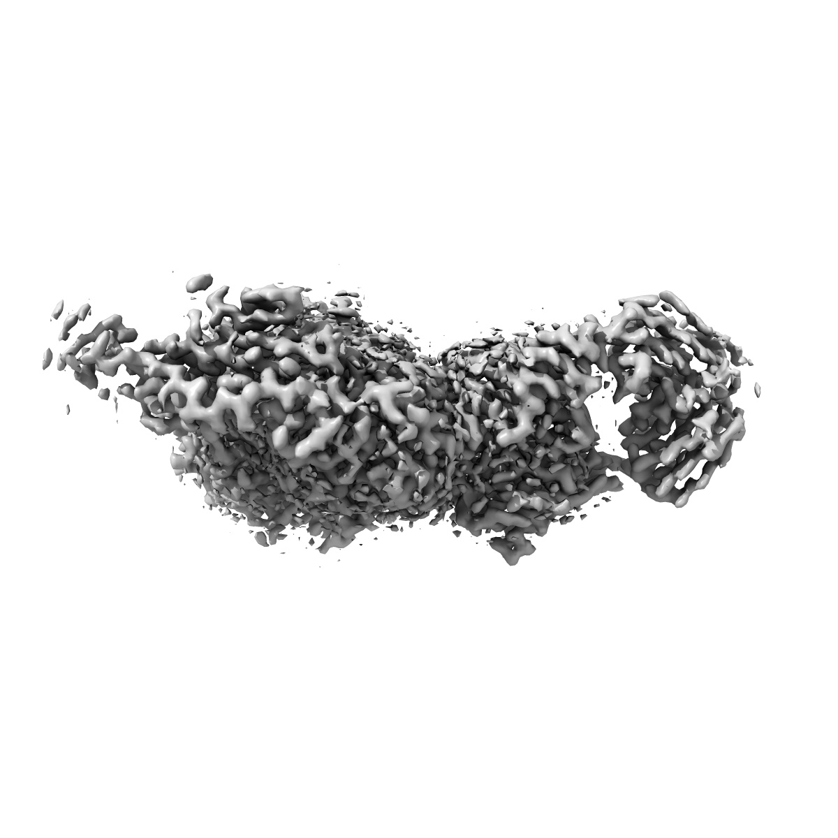
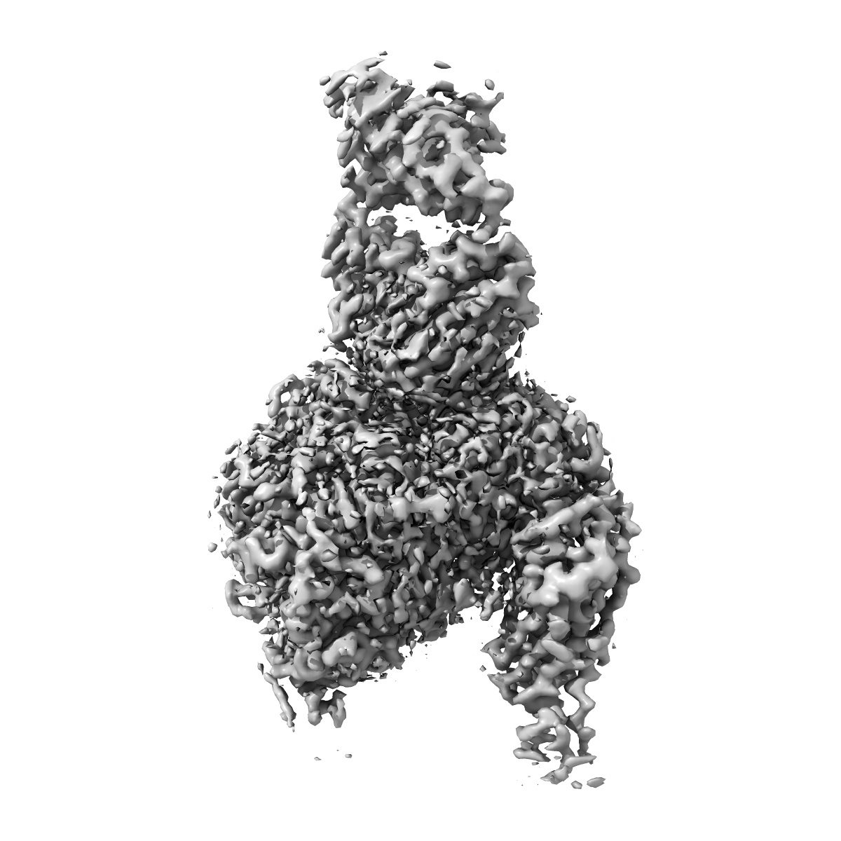
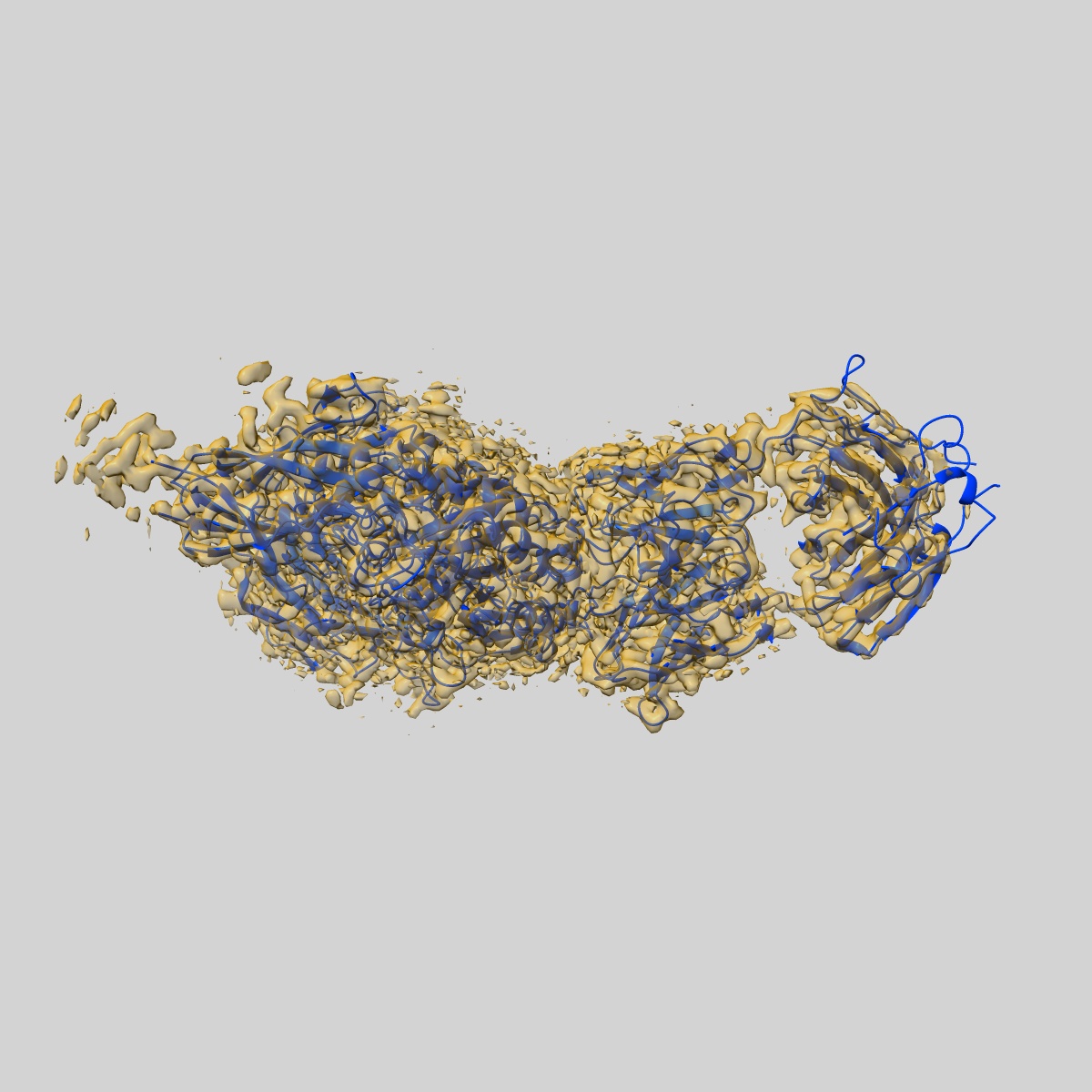
Cryo-EM structure of the integrin AlphaIIbBeta3-Abciximab complex

EMD-21044

Single-particle
2.8 Å
EMD-21044 Deposition: 28/11/2019
Map released: 05/02/2020
Last modified: 30/10/2024
Overview 3D View Sample Experiment Validation Volume Browser Additional data Links
Sample Organism: Homo sapiens, synthetic construct
Sample: Integrin AlphaIIbBeta3-Abciximab Complex
Fitted models: 6v4p (Avg. Q-score: 0.597)

Deposition Authors: Nesic D, Zhang Y
Cryo-Electron Microscopy Structure of the alpha IIb beta 3-Abciximab Complex.
Nesic D, Zhang Y, Spasic A, Li J, Provasi D, Filizola M, Walz T , Coller BS
(2020) Arterioscler Thromb Vasc Biol , 40 , 624 - 637
PUBMED: 31969014
DOI: doi:10.1161/ATVBAHA.119.313671
ISSN: 1524-4636
Abstract:
Objective
The αIIbβ3 antagonist antiplatelet drug abciximab is the chimeric antigen-binding fragment comprising the variable regions of murine monoclonal antibody 7E3 and the constant domains of human IgG1 and light chain κ. Previous mutagenesis studies suggested that abciximab binds to the β3 C177-C184 specificity-determining loop (SDL) and Trp129 on the adjacent β1-α1 helix. These studies could not, however, assess whether 7E3 or abciximab prevents fibrinogen binding by steric interference, disruption of either the αIIbβ3-binding pocket for fibrinogen or the β3 SDL (which is not part of the binding pocket but affects fibrinogen binding), or some combination of these effects. To address this gap, we used cryo-electron microscopy to determine the structure of the αIIbβ3-abciximab complex at 2.8 Å resolution. Approach and Results: The interacting surface of abciximab is comprised of residues from all 3 complementarity-determining regions of both the light and heavy chains, with high representation of aromatic residues. Binding is primarily to the β3 SDL and neighboring residues, the β1-α1 helix, and β3 residues Ser211, Val212 and Met335. Unexpectedly, the structure also indicated several interactions with αIIb. As judged by the cryo-electron microscopy model, molecular-dynamics simulations, and mutagenesis, the binding of abciximab does not appear to rely on the interaction with the αIIb residues and does not result in disruption of the fibrinogen-binding pocket; it does, however, compress and reduce the flexibility of the SDL.
Conclusions
We deduce that abciximab prevents ligand binding by steric interference, with a potential contribution via displacement of the SDL and limitation of the flexibility of the SDL residues.