EMD-21203
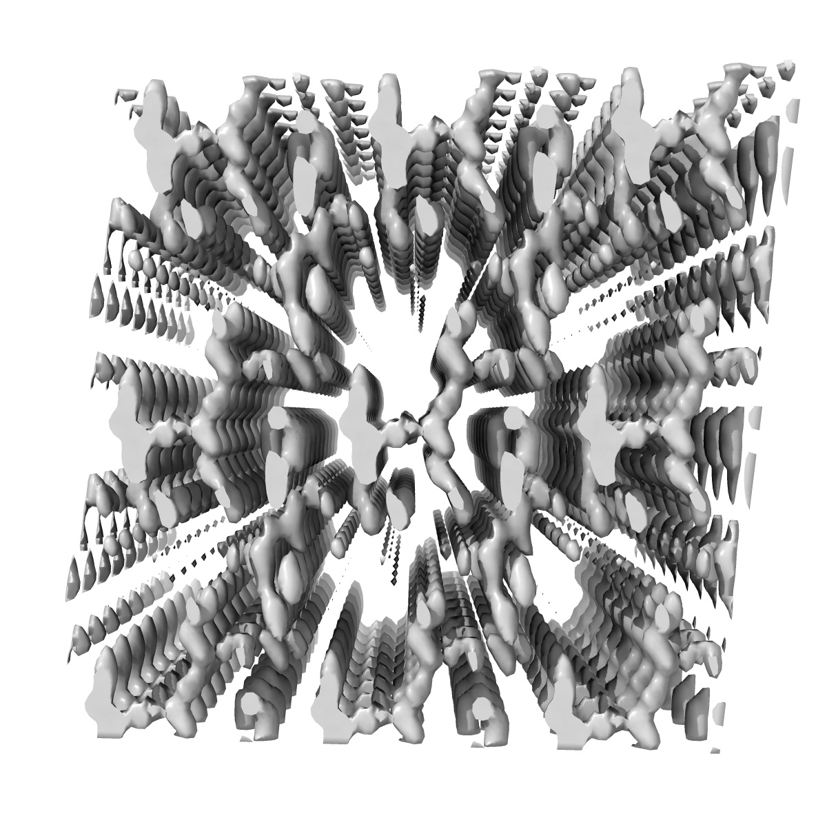
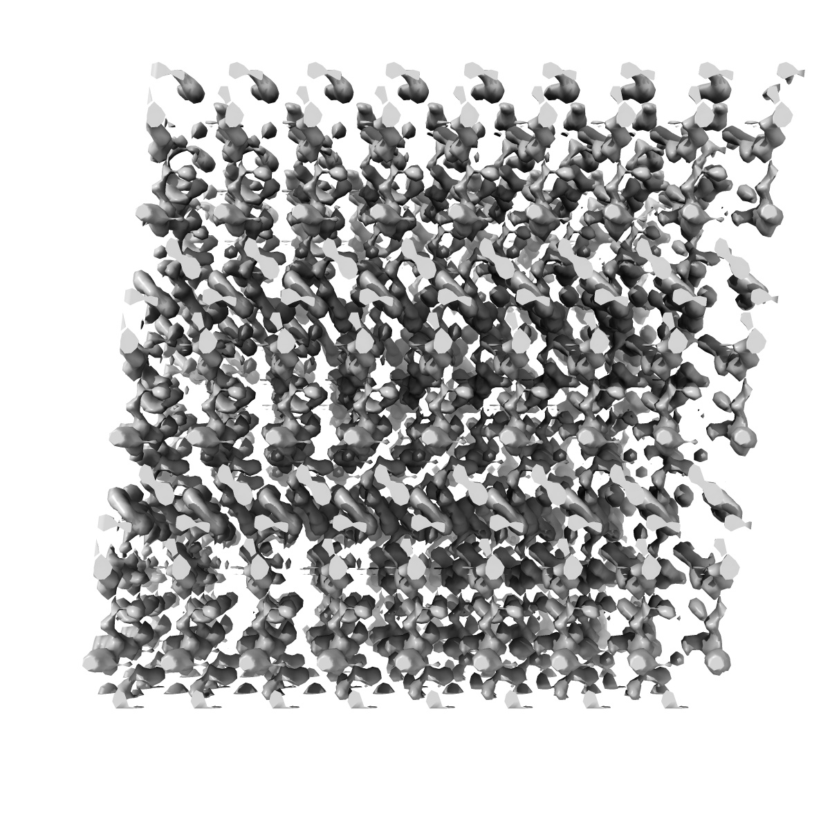
1.4A damaged structure of GSNQNNF used to determine initial phases from radiation damage
EMD-21203
Electron Crystallography Deposition: 09/01/2020
Map released: 19/02/2020
Last modified: 06/03/2024
Sample Organism:
synthetic construct
Sample: Synthetic proto-filament
Fitted models: 6vhc
Deposition Authors: Martynowycz MW, Hattne J
Sample: Synthetic proto-filament
Fitted models: 6vhc
Deposition Authors: Martynowycz MW, Hattne J

Experimental Phasing of MicroED Data Using Radiation Damage.
Abstract:
We previously demonstrated that microcrystal electron diffraction (MicroED) can be used to determine atomic-resolution structures from vanishingly small three-dimensional crystals. Here, we present an example of an experimentally phased structure using only MicroED data. The structure of a seven-residue peptide is solved starting from differences to the diffraction intensities induced by structural changes due to radiation damage. The same wedge of reciprocal space was recorded twice by continuous-rotation MicroED from a set of 11 individual crystals. The data from the first pass were merged to make a "low-dose dataset." The data from the second pass were similarly merged to form a "damaged dataset." Differences between these two datasets were used to identify a single heavy-atom site from a Patterson difference map, and initial phases were generated. Finally, the structure was completed by iterative cycles of modeling and refinement.
We previously demonstrated that microcrystal electron diffraction (MicroED) can be used to determine atomic-resolution structures from vanishingly small three-dimensional crystals. Here, we present an example of an experimentally phased structure using only MicroED data. The structure of a seven-residue peptide is solved starting from differences to the diffraction intensities induced by structural changes due to radiation damage. The same wedge of reciprocal space was recorded twice by continuous-rotation MicroED from a set of 11 individual crystals. The data from the first pass were merged to make a "low-dose dataset." The data from the second pass were similarly merged to form a "damaged dataset." Differences between these two datasets were used to identify a single heavy-atom site from a Patterson difference map, and initial phases were generated. Finally, the structure was completed by iterative cycles of modeling and refinement.