EMD-24663
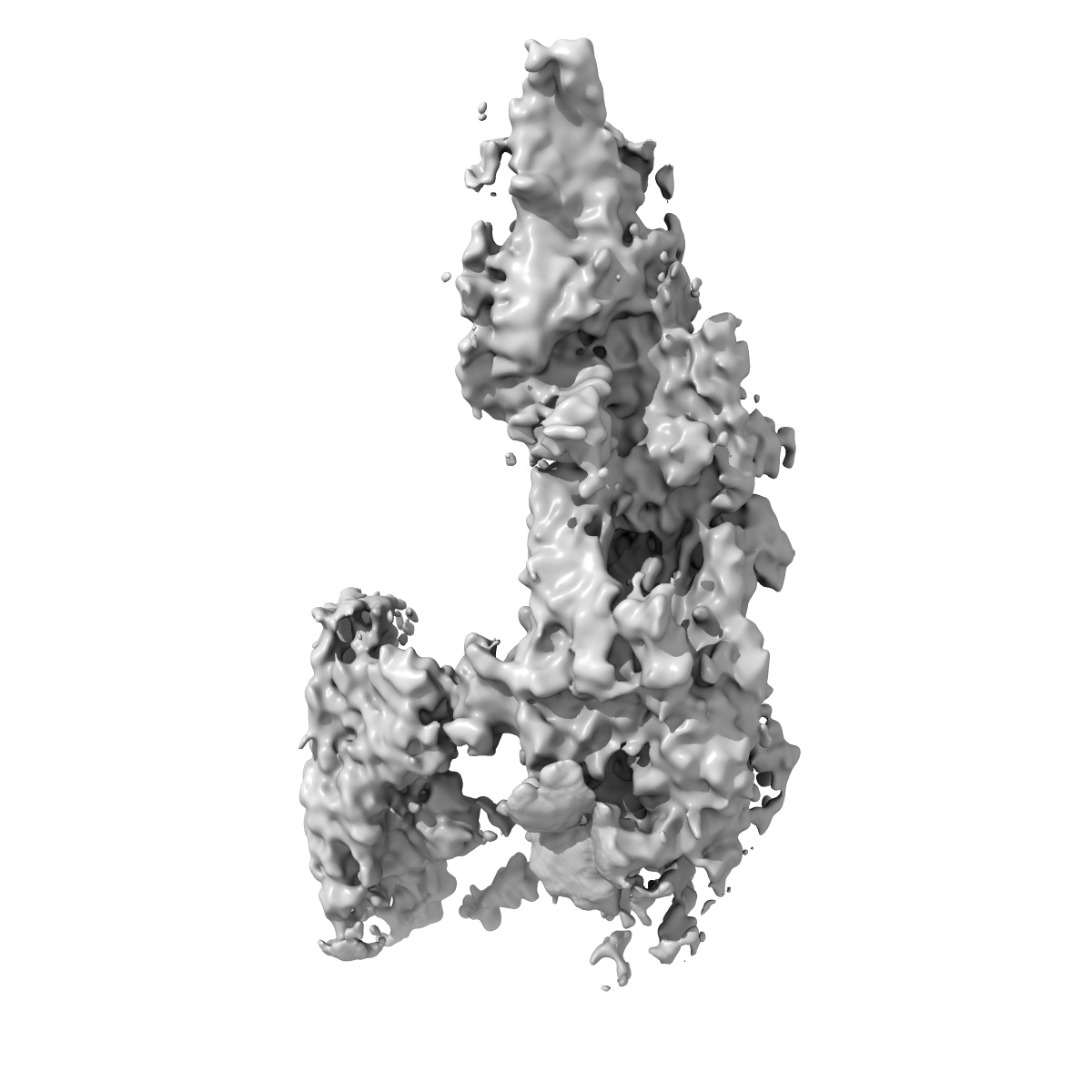
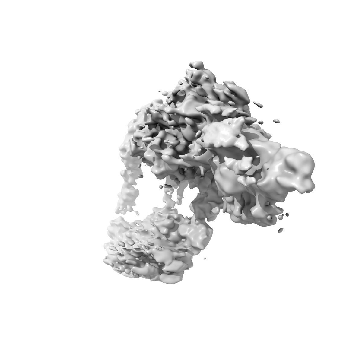
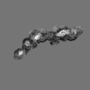
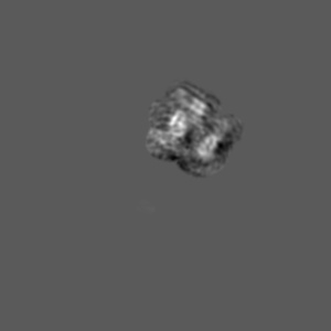
Cryo-EM density map of the outer dynein arm core from bovine tracheal cilia
EMD-24663
Single-particle8.0 Å
 Deposition: 10/08/2021
Map released: 27/10/2021
Last modified: 24/11/2021
Sample Organism:
Bos taurus
Sample: Outer dynein arm bound to doublet microtubule
Deposition Authors: Gui M ,
Anderson JR ,
Botsch JJ ,
Meleppattu S ,
Singh SK ,
Zhang Q,
Brown A
,
Anderson JR ,
Botsch JJ ,
Meleppattu S ,
Singh SK ,
Zhang Q,
Brown A
Sample: Outer dynein arm bound to doublet microtubule
Deposition Authors: Gui M
 ,
Anderson JR ,
Botsch JJ ,
Meleppattu S ,
Singh SK ,
Zhang Q,
Brown A
,
Anderson JR ,
Botsch JJ ,
Meleppattu S ,
Singh SK ,
Zhang Q,
Brown A
De novo identification of mammalian ciliary motility proteins using cryo-EM.
Gui M ,
Farley H ,
Anujan P,
Anderson JR ,
Maxwell DW ,
Whitchurch JB,
Botsch JJ ,
Qiu T ,
Meleppattu S ,
Singh SK ,
Zhang Q,
Thompson J ,
Lucas JS ,
Bingle CD ,
Norris DP,
Roy S,
Brown A
(2021) Cell , 184 , 5791 - 5806.e19
,
Farley H ,
Anujan P,
Anderson JR ,
Maxwell DW ,
Whitchurch JB,
Botsch JJ ,
Qiu T ,
Meleppattu S ,
Singh SK ,
Zhang Q,
Thompson J ,
Lucas JS ,
Bingle CD ,
Norris DP,
Roy S,
Brown A
(2021) Cell , 184 , 5791 - 5806.e19
Abstract:
Dynein-decorated doublet microtubules (DMTs) are critical components of the oscillatory molecular machine of cilia, the axoneme, and have luminal surfaces patterned periodically by microtubule inner proteins (MIPs). Here we present an atomic model of the 48-nm repeat of a mammalian DMT, derived from a cryoelectron microscopy (cryo-EM) map of the complex isolated from bovine respiratory cilia. The structure uncovers principles of doublet microtubule organization and features specific to vertebrate cilia, including previously unknown MIPs, a luminal bundle of tektin filaments, and a pentameric dynein-docking complex. We identify a mechanism for bridging 48- to 24-nm periodicity across the microtubule wall and show that loss of the proteins involved causes defective ciliary motility and laterality abnormalities in zebrafish and mice. Our structure identifies candidate genes for diagnosis of ciliopathies and provides a framework to understand their functions in driving ciliary motility.
Dynein-decorated doublet microtubules (DMTs) are critical components of the oscillatory molecular machine of cilia, the axoneme, and have luminal surfaces patterned periodically by microtubule inner proteins (MIPs). Here we present an atomic model of the 48-nm repeat of a mammalian DMT, derived from a cryoelectron microscopy (cryo-EM) map of the complex isolated from bovine respiratory cilia. The structure uncovers principles of doublet microtubule organization and features specific to vertebrate cilia, including previously unknown MIPs, a luminal bundle of tektin filaments, and a pentameric dynein-docking complex. We identify a mechanism for bridging 48- to 24-nm periodicity across the microtubule wall and show that loss of the proteins involved causes defective ciliary motility and laterality abnormalities in zebrafish and mice. Our structure identifies candidate genes for diagnosis of ciliopathies and provides a framework to understand their functions in driving ciliary motility.