EMD-27902
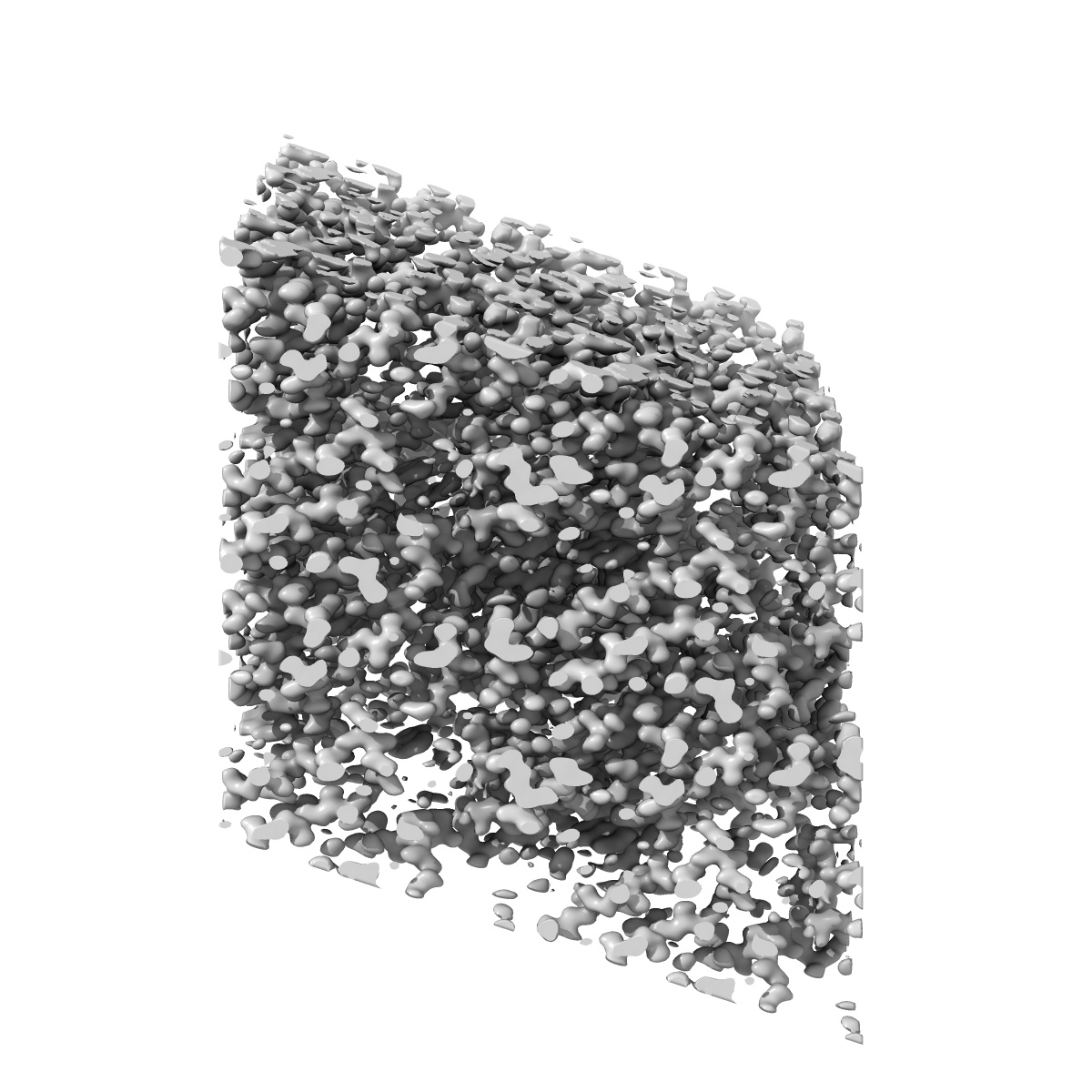
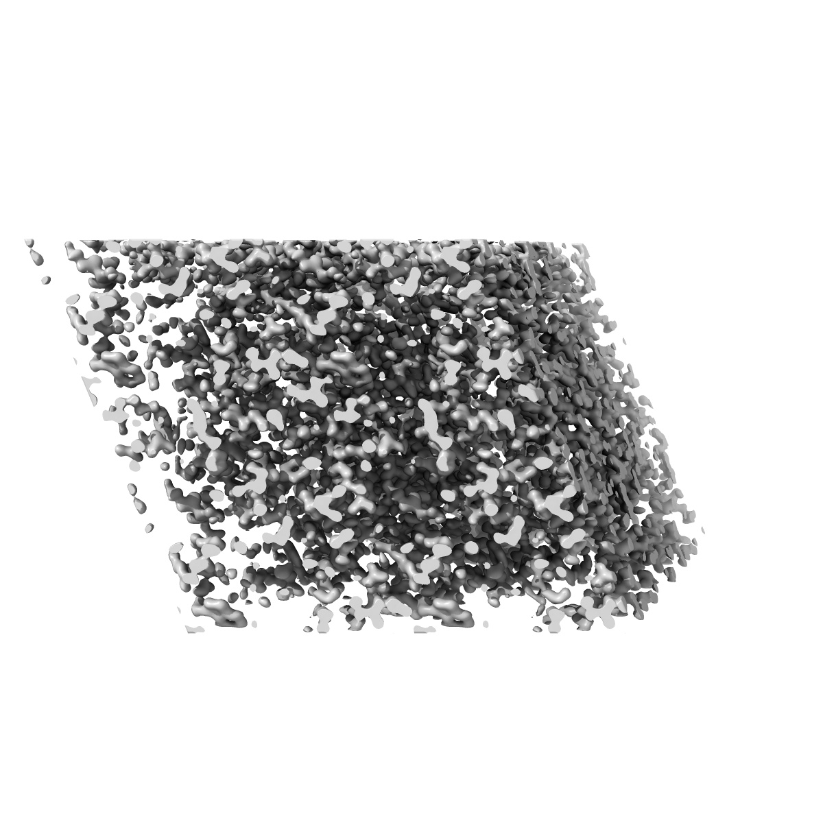
MicroED structure of triclinic lysozyme recorded on K3
EMD-27902
Electron Crystallography Deposition: 19/08/2022
Map released: 21/09/2022
Last modified: 23/10/2024
Sample Organism:
Gallus gallus
Sample: Lysozyme
Fitted models: 8e54
Deposition Authors: Clabbers MTB, Martynowycz MW, Hattne J ,
Nannenga BL,
Gonen T
,
Nannenga BL,
Gonen T
Sample: Lysozyme
Fitted models: 8e54
Deposition Authors: Clabbers MTB, Martynowycz MW, Hattne J
 ,
Nannenga BL,
Gonen T
,
Nannenga BL,
Gonen T
Electron-counting MicroED data with the K2 and K3 direct electron detectors.
Clabbers MTB,
Martynowycz MW,
Hattne J ,
Nannenga BL,
Gonen T
(2022) J Struct Biol , 214 , 107886 - 107886
,
Nannenga BL,
Gonen T
(2022) J Struct Biol , 214 , 107886 - 107886
Abstract:
Microcrystal electron diffraction (MicroED) uses electron cryo-microscopy (cryo-EM) to collect diffraction data from small crystals during continuous rotation of the sample. As a result of advances in hardware as well as methods development, the data quality has continuously improved over the past decade, to the point where even macromolecular structures can be determined ab initio. Detectors suitable for electron diffraction should ideally have fast readout to record data in movie mode, and high sensitivity at low exposure rates to accurately report the intensities. Direct electron detectors are commonly used in cryo-EM imaging for their sensitivity and speed, but despite their availability are generally not used in diffraction. Primary concerns with diffraction experiments are the dynamic range and coincidence loss, which will corrupt the measurement if the flux exceeds the count rate of the detector. Here, we describe instrument setup and low-exposure MicroED data collection in electron-counting mode using K2 and K3 direct electron detectors and show that the integrated intensities can be effectively used to solve structures of two macromolecules between 1.2 Å and 2.8 Å resolution. Even though a beam stop was not used with the K3 studies we did not observe damage to the camera. As these cameras are already available in many cryo-EM facilities, this provides opportunities for users who do not have access to dedicated facilities for MicroED.
Microcrystal electron diffraction (MicroED) uses electron cryo-microscopy (cryo-EM) to collect diffraction data from small crystals during continuous rotation of the sample. As a result of advances in hardware as well as methods development, the data quality has continuously improved over the past decade, to the point where even macromolecular structures can be determined ab initio. Detectors suitable for electron diffraction should ideally have fast readout to record data in movie mode, and high sensitivity at low exposure rates to accurately report the intensities. Direct electron detectors are commonly used in cryo-EM imaging for their sensitivity and speed, but despite their availability are generally not used in diffraction. Primary concerns with diffraction experiments are the dynamic range and coincidence loss, which will corrupt the measurement if the flux exceeds the count rate of the detector. Here, we describe instrument setup and low-exposure MicroED data collection in electron-counting mode using K2 and K3 direct electron detectors and show that the integrated intensities can be effectively used to solve structures of two macromolecules between 1.2 Å and 2.8 Å resolution. Even though a beam stop was not used with the K3 studies we did not observe damage to the camera. As these cameras are already available in many cryo-EM facilities, this provides opportunities for users who do not have access to dedicated facilities for MicroED.