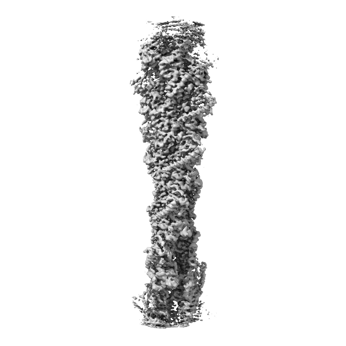
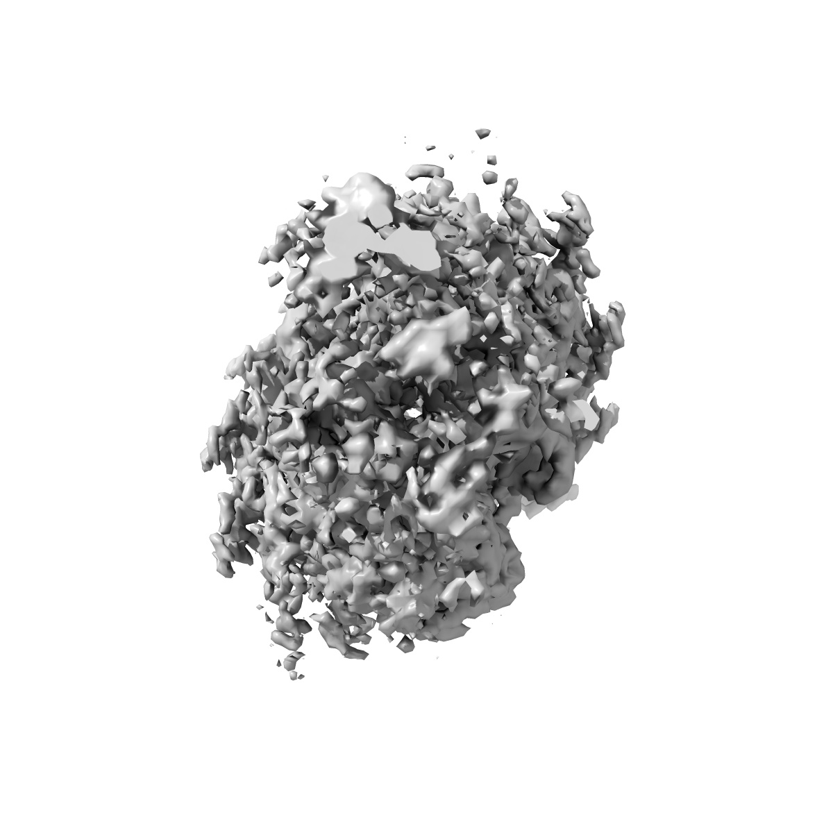
EMD-30085
F-actin-Utrophin complex
EMD-30085
Helical reconstruction3.6 Å
 Deposition: 10/03/2020
Map released: 20/05/2020
Last modified: 27/03/2024
Sample Organism:
Gallus gallus,
Homo sapiens
Sample: Filamentous actin in ADP state
Fitted models: 6m5g (Avg. Q-score: 0.53)
Deposition Authors: Kumari A, Ragunath VK
Sample: Filamentous actin in ADP state
Fitted models: 6m5g (Avg. Q-score: 0.53)
Deposition Authors: Kumari A, Ragunath VK
Structural insights into actin filament recognition by commonly used cellular actin markers.
Kumari A,
Kesarwani S  ,
Javoor MG ,
Vinothkumar KR,
Sirajuddin M
,
Javoor MG ,
Vinothkumar KR,
Sirajuddin M
(2020) EMBO J , 39 , e104006 - e104006
 ,
Javoor MG ,
Vinothkumar KR,
Sirajuddin M
,
Javoor MG ,
Vinothkumar KR,
Sirajuddin M
(2020) EMBO J , 39 , e104006 - e104006
Abstract:
Cellular studies of filamentous actin (F-actin) processes commonly utilize fluorescent versions of toxins, peptides, and proteins that bind actin. While the choice of these markers has been largely based on availability and ease, there is a severe dearth of structural data for an informed judgment in employing suitable F-actin markers for a particular requirement. Here, we describe the electron cryomicroscopy structures of phalloidin, lifeAct, and utrophin bound to F-actin, providing a comprehensive high-resolution structural comparison of widely used actin markers and their influence towards F-actin. Our results show that phalloidin binding does not induce specific conformational change and lifeAct specifically recognizes closed D-loop conformation, i.e., ADP-Pi or ADP states of F-actin. The structural models aided designing of minimal utrophin and a shorter lifeAct, which can be utilized as F-actin marker. Together, our study provides a structural perspective, where the binding sites of utrophin and lifeAct overlap with majority of actin-binding proteins and thus offering an invaluable resource for researchers in choosing appropriate actin markers and generating new marker variants.
Cellular studies of filamentous actin (F-actin) processes commonly utilize fluorescent versions of toxins, peptides, and proteins that bind actin. While the choice of these markers has been largely based on availability and ease, there is a severe dearth of structural data for an informed judgment in employing suitable F-actin markers for a particular requirement. Here, we describe the electron cryomicroscopy structures of phalloidin, lifeAct, and utrophin bound to F-actin, providing a comprehensive high-resolution structural comparison of widely used actin markers and their influence towards F-actin. Our results show that phalloidin binding does not induce specific conformational change and lifeAct specifically recognizes closed D-loop conformation, i.e., ADP-Pi or ADP states of F-actin. The structural models aided designing of minimal utrophin and a shorter lifeAct, which can be utilized as F-actin marker. Together, our study provides a structural perspective, where the binding sites of utrophin and lifeAct overlap with majority of actin-binding proteins and thus offering an invaluable resource for researchers in choosing appropriate actin markers and generating new marker variants.