EMD-3244
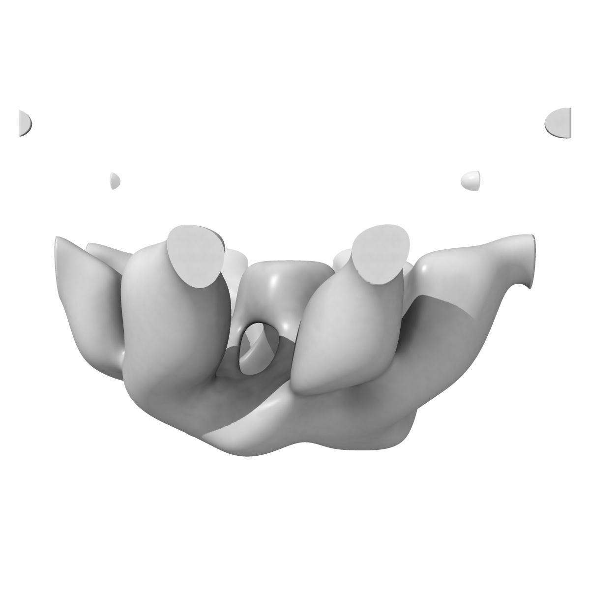
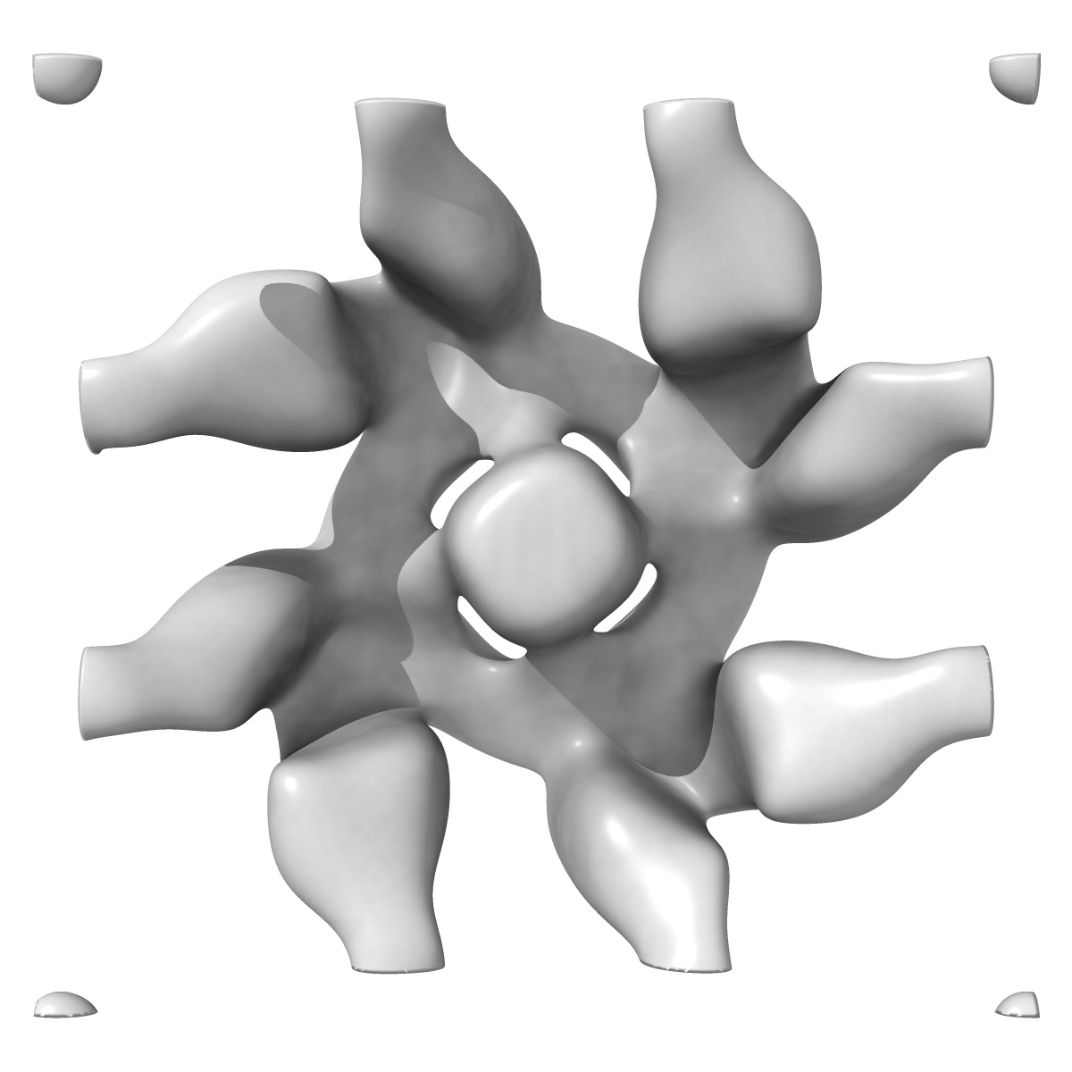
Electron negative-staining microscopy of an aerolysin-like protein
EMD-3244
Electron Crystallography20.0 Å
 Deposition: 13/11/2015
Map released: 13/01/2016
Last modified: 09/03/2016
Sample Organism:
Danio rerio
Sample: Dln1 (a zebrafish aerolysin-like protein) oligomer on phospholipid monolayer
Deposition Authors: Jia N, Liu N, Cheng W, Jiang YL, Sun H, Chen LL, Peng JH, Zhang YH, Zhang ZH, Wang XJ, Cai G, Wang JF, Zhang ZY, Wu H, Wang HW, Chen YX, Zhou CZ
Sample: Dln1 (a zebrafish aerolysin-like protein) oligomer on phospholipid monolayer
Deposition Authors: Jia N, Liu N, Cheng W, Jiang YL, Sun H, Chen LL, Peng JH, Zhang YH, Zhang ZH, Wang XJ, Cai G, Wang JF, Zhang ZY, Wu H, Wang HW, Chen YX, Zhou CZ
Structural basis for receptor recognition and pore formation of a zebrafish aerolysin-like protein
Jia N  ,
Liu N ,
Cheng W,
Jiang YL ,
Sun H,
Chen LL,
Peng JH ,
Zhang YH,
Zhang ZH ,
Wang XJ,
Cai G,
Wang JF ,
Zhang ZY,
Wu H,
Wang HW,
Chen YX ,
Zhou CZ
,
Liu N ,
Cheng W,
Jiang YL ,
Sun H,
Chen LL,
Peng JH ,
Zhang YH,
Zhang ZH ,
Wang XJ,
Cai G,
Wang JF ,
Zhang ZY,
Wu H,
Wang HW,
Chen YX ,
Zhou CZ
(2016) Embo Rep. , 17 , 235 - 248
 ,
Liu N ,
Cheng W,
Jiang YL ,
Sun H,
Chen LL,
Peng JH ,
Zhang YH,
Zhang ZH ,
Wang XJ,
Cai G,
Wang JF ,
Zhang ZY,
Wu H,
Wang HW,
Chen YX ,
Zhou CZ
,
Liu N ,
Cheng W,
Jiang YL ,
Sun H,
Chen LL,
Peng JH ,
Zhang YH,
Zhang ZH ,
Wang XJ,
Cai G,
Wang JF ,
Zhang ZY,
Wu H,
Wang HW,
Chen YX ,
Zhou CZ
(2016) Embo Rep. , 17 , 235 - 248
Abstract:
Various aerolysin-like pore-forming proteins have been identified from bacteria to vertebrates. However, the mechanism of receptor recognition and/or pore formation of the eukaryotic members remains unknown. Here, we present the first crystal and electron microscopy structures of a vertebrate aerolysin-like protein from Danio rerio, termed Dln1, before and after pore formation. Each subunit of Dln1 dimer comprises a β-prism lectin module followed by an aerolysin module. Specific binding of the lectin module toward high-mannose glycans triggers drastic conformational changes of the aerolysin module in a pH-dependent manner, ultimately resulting in the formation of a membrane-bound octameric pore. Structural analyses combined with computational simulations and biochemical assays suggest a pore-forming process with an activation mechanism distinct from the previously characterized bacterial members. Moreover, Dln1 and its homologs are ubiquitously distributed in bony fishes and lamprey, suggesting a novel fish-specific defense molecule.
Various aerolysin-like pore-forming proteins have been identified from bacteria to vertebrates. However, the mechanism of receptor recognition and/or pore formation of the eukaryotic members remains unknown. Here, we present the first crystal and electron microscopy structures of a vertebrate aerolysin-like protein from Danio rerio, termed Dln1, before and after pore formation. Each subunit of Dln1 dimer comprises a β-prism lectin module followed by an aerolysin module. Specific binding of the lectin module toward high-mannose glycans triggers drastic conformational changes of the aerolysin module in a pH-dependent manner, ultimately resulting in the formation of a membrane-bound octameric pore. Structural analyses combined with computational simulations and biochemical assays suggest a pore-forming process with an activation mechanism distinct from the previously characterized bacterial members. Moreover, Dln1 and its homologs are ubiquitously distributed in bony fishes and lamprey, suggesting a novel fish-specific defense molecule.