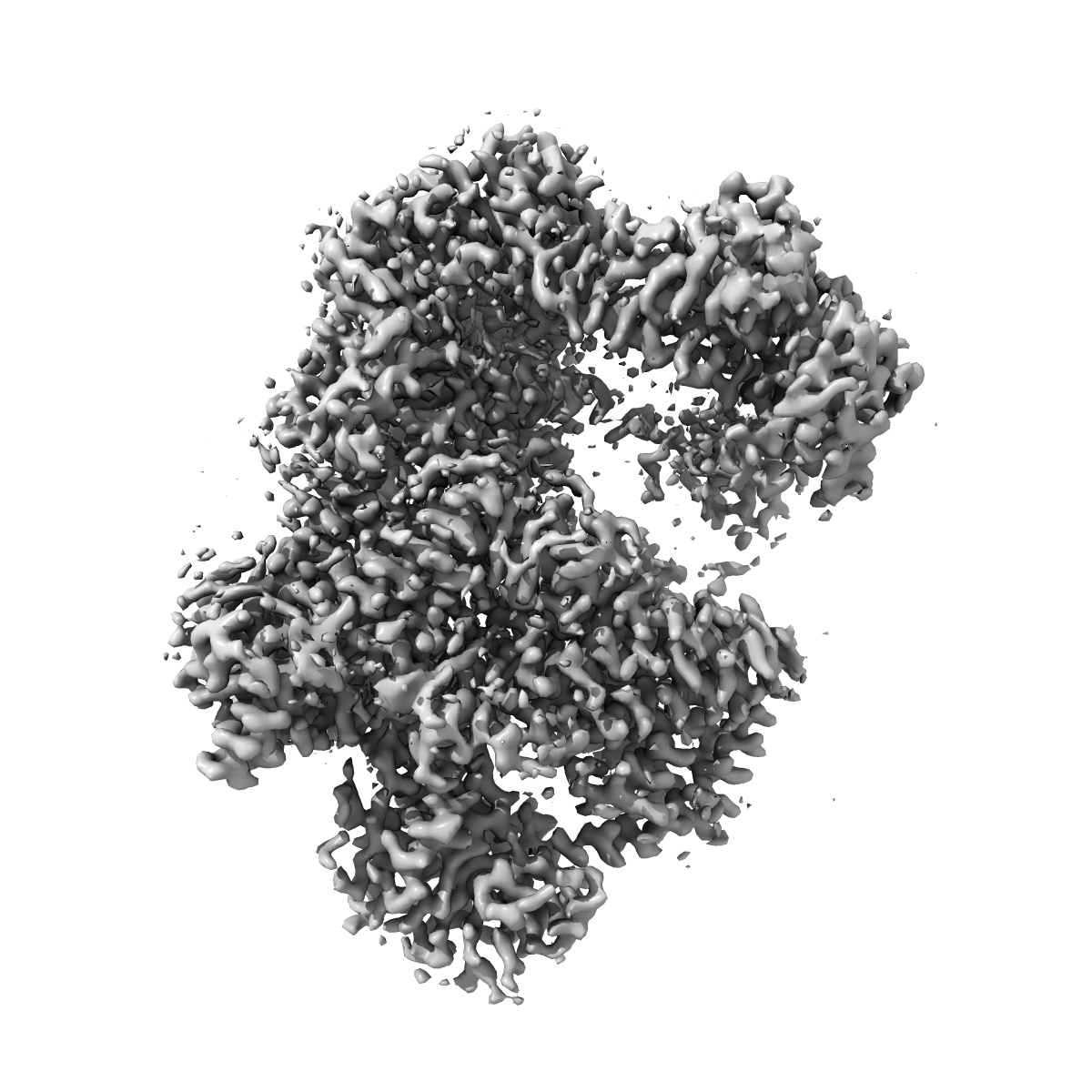
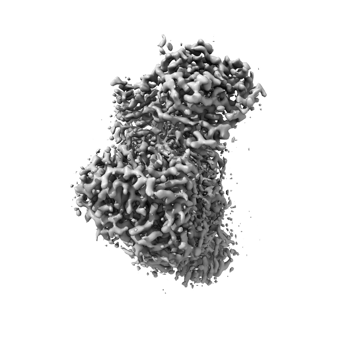
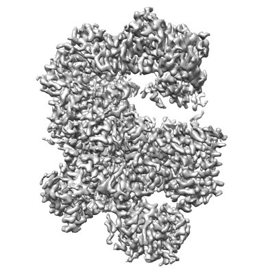
EMD-33243
Cryo-EM structure of Na+-pumping NADH-ubiquinone oxidoreductase from Vibrio cholerae, state 2
EMD-33243
Single-particle3.1 Å
 Deposition: 19/04/2022
Map released: 20/07/2022
Last modified: 10/08/2022
Sample Organism:
Vibrio cholerae O395
Sample: Na+-pumping NADH-ubiquinone oxidoreductase from Vibrio cholerae, state 2
Fitted models: 7xk4 (Avg. Q-score: 0.562)
Raw data: EMPIAR-11210
Deposition Authors: Kishikawa J ,
Ishikawa M ,
Masuya T ,
Murai M ,
Barquera B ,
Miyoshi H
,
Ishikawa M ,
Masuya T ,
Murai M ,
Barquera B ,
Miyoshi H
Sample: Na+-pumping NADH-ubiquinone oxidoreductase from Vibrio cholerae, state 2
Fitted models: 7xk4 (Avg. Q-score: 0.562)
Raw data: EMPIAR-11210
Deposition Authors: Kishikawa J
 ,
Ishikawa M ,
Masuya T ,
Murai M ,
Barquera B ,
Miyoshi H
,
Ishikawa M ,
Masuya T ,
Murai M ,
Barquera B ,
Miyoshi H
Cryo-EM structures of Na + -pumping NADH-ubiquinone oxidoreductase from Vibrio cholerae.
Kishikawa JI ,
Ishikawa M ,
Masuya T ,
Murai M ,
Kitazumi Y,
Butler NL,
Kato T ,
Barquera B ,
Miyoshi H
(2022) Nat Commun , 13 , 4082 - 4082
,
Ishikawa M ,
Masuya T ,
Murai M ,
Kitazumi Y,
Butler NL,
Kato T ,
Barquera B ,
Miyoshi H
(2022) Nat Commun , 13 , 4082 - 4082
Abstract:
The Na+-pumping NADH-ubiquinone oxidoreductase (Na+-NQR) couples electron transfer from NADH to ubiquinone with Na+-pumping, generating an electrochemical Na+ gradient that is essential for energy-consuming reactions in bacteria. Since Na+-NQR is exclusively found in prokaryotes, it is a promising target for highly selective antibiotics. However, the molecular mechanism of inhibition is not well-understood for lack of the atomic structural information about an inhibitor-bound state. Here we present cryo-electron microscopy structures of Na+-NQR from Vibrio cholerae with or without a bound inhibitor at 2.5- to 3.1-Å resolution. The structures reveal the arrangement of all six redox cofactors including a herein identified 2Fe-2S cluster located between the NqrD and NqrE subunits. A large part of the hydrophilic NqrF is barely visible in the density map, suggesting a high degree of flexibility. This flexibility may be responsible to reducing the long distance between the 2Fe-2S centers in NqrF and NqrD/E. Two different types of specific inhibitors bind to the N-terminal region of NqrB, which is disordered in the absence of inhibitors. The present study provides a foundation for understanding the function of Na+-NQR and the binding manner of specific inhibitors.
The Na+-pumping NADH-ubiquinone oxidoreductase (Na+-NQR) couples electron transfer from NADH to ubiquinone with Na+-pumping, generating an electrochemical Na+ gradient that is essential for energy-consuming reactions in bacteria. Since Na+-NQR is exclusively found in prokaryotes, it is a promising target for highly selective antibiotics. However, the molecular mechanism of inhibition is not well-understood for lack of the atomic structural information about an inhibitor-bound state. Here we present cryo-electron microscopy structures of Na+-NQR from Vibrio cholerae with or without a bound inhibitor at 2.5- to 3.1-Å resolution. The structures reveal the arrangement of all six redox cofactors including a herein identified 2Fe-2S cluster located between the NqrD and NqrE subunits. A large part of the hydrophilic NqrF is barely visible in the density map, suggesting a high degree of flexibility. This flexibility may be responsible to reducing the long distance between the 2Fe-2S centers in NqrF and NqrD/E. Two different types of specific inhibitors bind to the N-terminal region of NqrB, which is disordered in the absence of inhibitors. The present study provides a foundation for understanding the function of Na+-NQR and the binding manner of specific inhibitors.