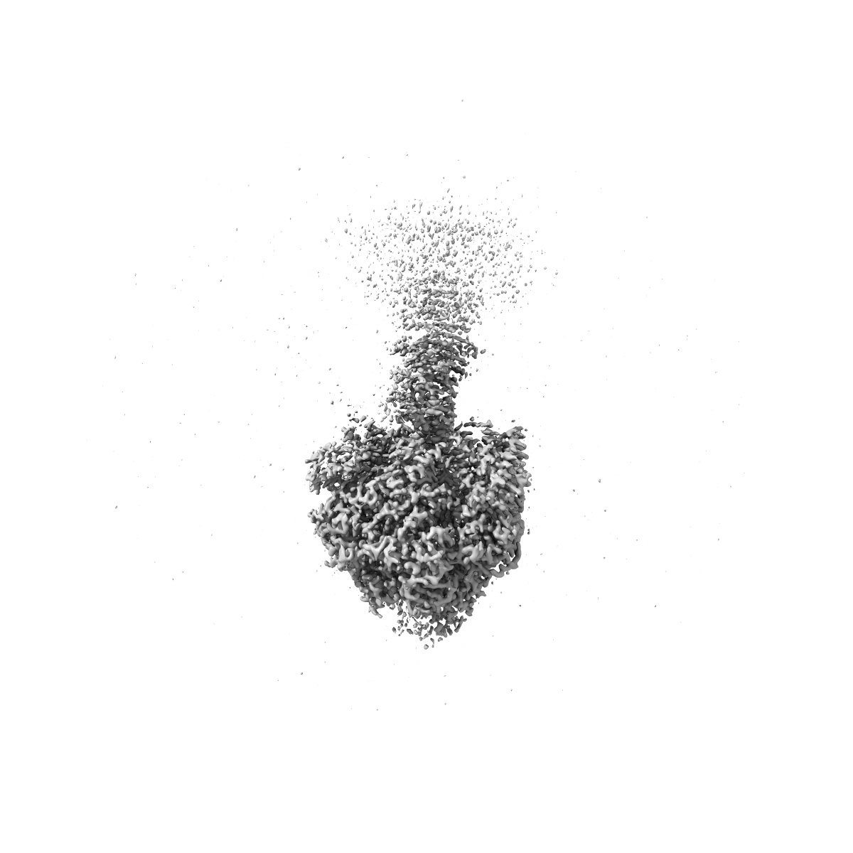
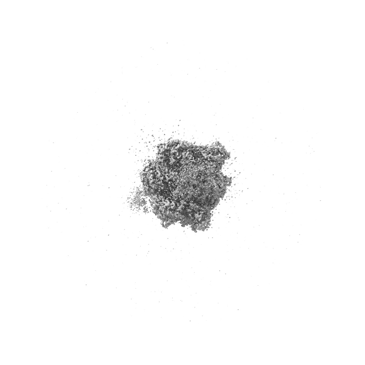
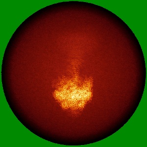
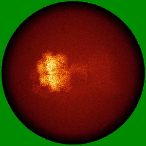
EMD-34572
Human ATP synthase F1 domain, state 3a
EMD-34572
Single-particle2.61 Å
 Deposition: 25/10/2022
Map released: 31/05/2023
Last modified: 03/07/2024
Sample Organism:
Homo sapiens
Sample: Human ATP synthase
Fitted models: 8h9l (Avg. Q-score: 0.615)
Deposition Authors: Lai Y ,
Zhang Y,
Liu F ,
Gao Y,
Gong H ,
Rao Z
,
Zhang Y,
Liu F ,
Gao Y,
Gong H ,
Rao Z
Sample: Human ATP synthase
Fitted models: 8h9l (Avg. Q-score: 0.615)
Deposition Authors: Lai Y
 ,
Zhang Y,
Liu F ,
Gao Y,
Gong H ,
Rao Z
,
Zhang Y,
Liu F ,
Gao Y,
Gong H ,
Rao Z
Structure of the human ATP synthase.
Lai Y ,
Zhang Y,
Zhou S,
Xu J,
Du Z,
Feng Z,
Yu L,
Zhao Z,
Wang W,
Tang Y,
Yang X,
Guddat LW,
Liu F ,
Gao Y,
Rao Z,
Gong H
(2023) Mol Cell , 83 , 2137
,
Zhang Y,
Zhou S,
Xu J,
Du Z,
Feng Z,
Yu L,
Zhao Z,
Wang W,
Tang Y,
Yang X,
Guddat LW,
Liu F ,
Gao Y,
Rao Z,
Gong H
(2023) Mol Cell , 83 , 2137
Abstract:
Biological energy currency ATP is produced by F1Fo-ATP synthase. However, the molecular mechanism for human ATP synthase action remains unknown. Here, we present snapshot images for three main rotational states and one substate of human ATP synthase using cryoelectron microscopy. These structures reveal that the release of ADP occurs when the β subunit of F1Fo-ATP synthase is in the open conformation, showing how ADP binding is coordinated during synthesis. The accommodation of the symmetry mismatch between F1 and Fo motors is resolved by the torsional flexing of the entire complex, especially the γ subunit, and the rotational substep of the c subunit. Water molecules are identified in the inlet and outlet half-channels, suggesting that the proton transfer in these two half-channels proceed via a Grotthus mechanism. Clinically relevant mutations are mapped to the structure, showing that they are mainly located at the subunit-subunit interfaces, thus causing instability of the complex.
Biological energy currency ATP is produced by F1Fo-ATP synthase. However, the molecular mechanism for human ATP synthase action remains unknown. Here, we present snapshot images for three main rotational states and one substate of human ATP synthase using cryoelectron microscopy. These structures reveal that the release of ADP occurs when the β subunit of F1Fo-ATP synthase is in the open conformation, showing how ADP binding is coordinated during synthesis. The accommodation of the symmetry mismatch between F1 and Fo motors is resolved by the torsional flexing of the entire complex, especially the γ subunit, and the rotational substep of the c subunit. Water molecules are identified in the inlet and outlet half-channels, suggesting that the proton transfer in these two half-channels proceed via a Grotthus mechanism. Clinically relevant mutations are mapped to the structure, showing that they are mainly located at the subunit-subunit interfaces, thus causing instability of the complex.