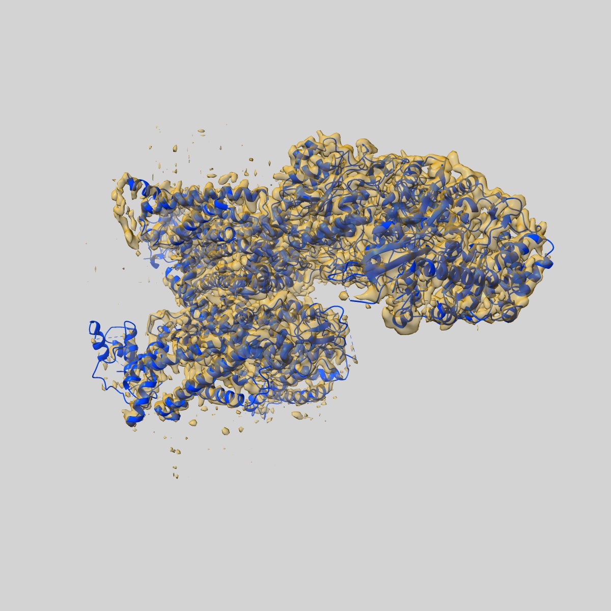
EMD-34891
Cryo-EM structure of human high-voltage activated L-type calcium channel CaV1.2 in complex with tetrandrine (TET)
EMD-34891
Single-particle3.4 Å
 Deposition: 02/12/2022
Map released: 24/04/2024
Last modified: 06/11/2024
Sample Organism:
Homo sapiens
Sample: cav1.2alpha2delta1beta2b
Fitted models: 8hma (Avg. Q-score: 0.424)
Deposition Authors: Wei Y ,
Yu Z,
Zhao Y
,
Yu Z,
Zhao Y
Sample: cav1.2alpha2delta1beta2b
Fitted models: 8hma (Avg. Q-score: 0.424)
Deposition Authors: Wei Y
 ,
Yu Z,
Zhao Y
,
Yu Z,
Zhao Y
Structural bases of inhibitory mechanism of Ca V 1.2 channel inhibitors.
Wei Y ,
Yu Z,
Wang L,
Li X,
Li N,
Bai Q,
Wang Y,
Li R,
Meng Y ,
Xu H,
Wang X,
Dong Y ,
Huang Z ,
Zhang XC ,
Zhao Y
(2024) Nat Commun , 15 , 2772 - 2772
,
Yu Z,
Wang L,
Li X,
Li N,
Bai Q,
Wang Y,
Li R,
Meng Y ,
Xu H,
Wang X,
Dong Y ,
Huang Z ,
Zhang XC ,
Zhao Y
(2024) Nat Commun , 15 , 2772 - 2772
Abstract:
The voltage-gated calcium channel CaV1.2 is essential for cardiac and vessel smooth muscle contractility and brain function. Accumulating evidence demonstrates that malfunctions of CaV1.2 are involved in brain and heart diseases. Pharmacological inhibition of CaV1.2 is therefore of therapeutic value. Here, we report cryo-EM structures of CaV1.2 in the absence or presence of the antirheumatic drug tetrandrine or antihypertensive drug benidipine. Tetrandrine acts as a pore blocker in a pocket composed of S6II, S6III, and S6IV helices and forms extensive hydrophobic interactions with CaV1.2. Our structure elucidates that benidipine is located in the DIII-DIV fenestration site. Its hydrophobic sidechain, phenylpiperidine, is positioned at the exterior of the pore domain and cradled within a hydrophobic pocket formed by S5DIII, S6DIII, and S6DIV helices, providing additional interactions to exert inhibitory effects on both L-type and T-type voltage gated calcium channels. These findings provide the structural foundation for the rational design and optimization of therapeutic inhibitors of voltage-gated calcium channels.
The voltage-gated calcium channel CaV1.2 is essential for cardiac and vessel smooth muscle contractility and brain function. Accumulating evidence demonstrates that malfunctions of CaV1.2 are involved in brain and heart diseases. Pharmacological inhibition of CaV1.2 is therefore of therapeutic value. Here, we report cryo-EM structures of CaV1.2 in the absence or presence of the antirheumatic drug tetrandrine or antihypertensive drug benidipine. Tetrandrine acts as a pore blocker in a pocket composed of S6II, S6III, and S6IV helices and forms extensive hydrophobic interactions with CaV1.2. Our structure elucidates that benidipine is located in the DIII-DIV fenestration site. Its hydrophobic sidechain, phenylpiperidine, is positioned at the exterior of the pore domain and cradled within a hydrophobic pocket formed by S5DIII, S6DIII, and S6DIV helices, providing additional interactions to exert inhibitory effects on both L-type and T-type voltage gated calcium channels. These findings provide the structural foundation for the rational design and optimization of therapeutic inhibitors of voltage-gated calcium channels.