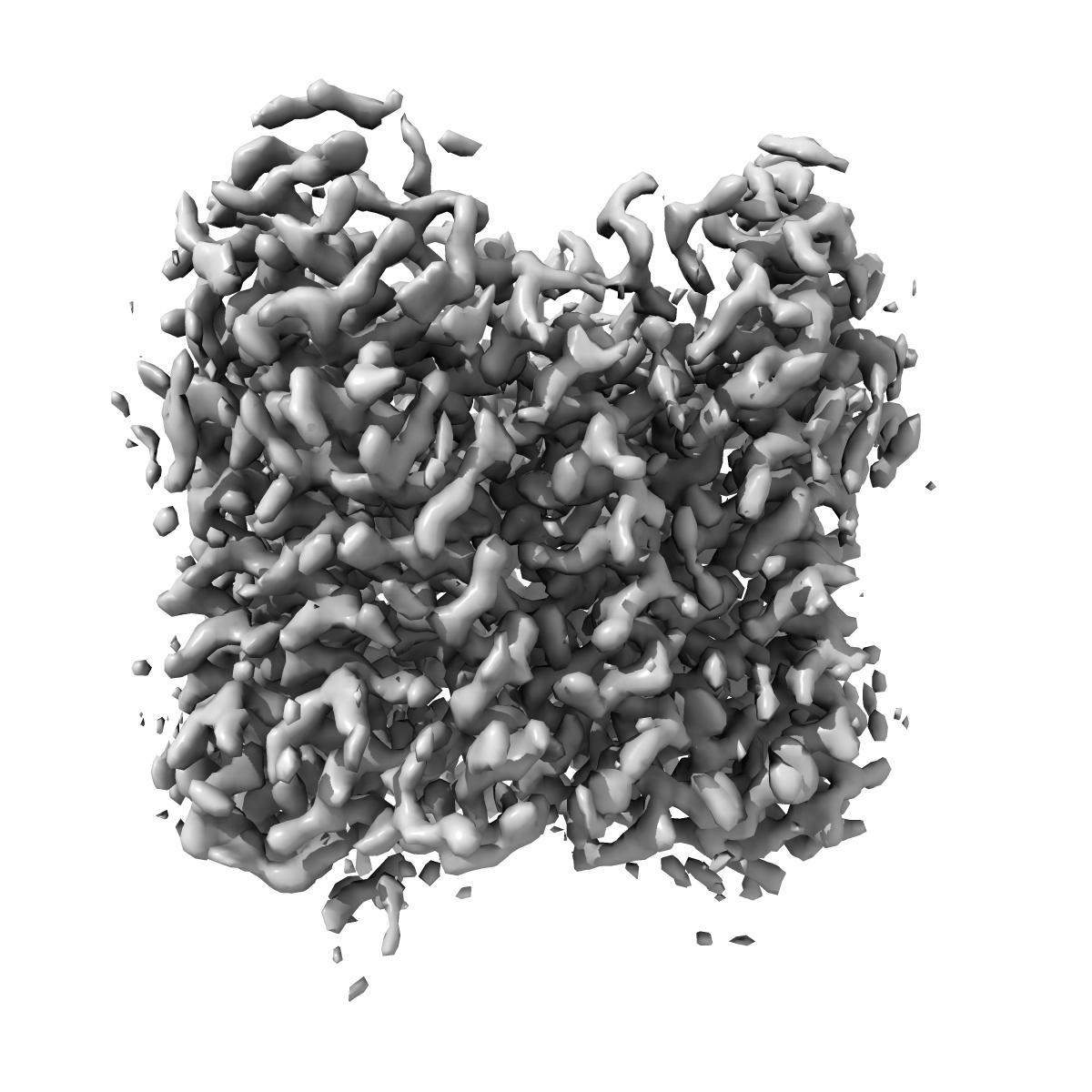
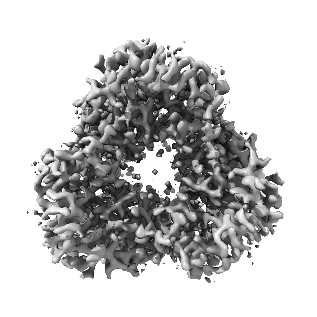
EMD-35713
Cryo-EM structure of the potassium-selective channelrhodopsin HcKCR1 H225F mutant in lipid nanodisc
EMD-35713
Single-particle2.66 Å
 Deposition: 23/03/2023
Map released: 06/09/2023
Last modified: 06/11/2024
Sample Organism:
Hyphochytrium catenoides
Sample: HcKCR1
Fitted models: 8iu0
Deposition Authors: Tajima S ,
Kim Y,
Nakamura S,
Yamashita K ,
Fukuda M,
Deisseroth K,
Kato HE
,
Kim Y,
Nakamura S,
Yamashita K ,
Fukuda M,
Deisseroth K,
Kato HE
Sample: HcKCR1
Fitted models: 8iu0
Deposition Authors: Tajima S
 ,
Kim Y,
Nakamura S,
Yamashita K ,
Fukuda M,
Deisseroth K,
Kato HE
,
Kim Y,
Nakamura S,
Yamashita K ,
Fukuda M,
Deisseroth K,
Kato HE
Structural basis for ion selectivity in potassium-selective channelrhodopsins.
Tajima S ,
Kim YS,
Fukuda M,
Jo Y ,
Wang PY ,
Paggi JM,
Inoue M,
Byrne EFX,
Kishi KE,
Nakamura S,
Ramakrishnan C,
Takaramoto S ,
Nagata T,
Konno M,
Sugiura M,
Katayama K,
Matsui TE,
Yamashita K ,
Kim S,
Ikeda H,
Kim J,
Kandori H,
Dror RO ,
Inoue K,
Deisseroth K,
Kato HE
(2023) Cell , 186 , 4325 - 4344.e26
,
Kim YS,
Fukuda M,
Jo Y ,
Wang PY ,
Paggi JM,
Inoue M,
Byrne EFX,
Kishi KE,
Nakamura S,
Ramakrishnan C,
Takaramoto S ,
Nagata T,
Konno M,
Sugiura M,
Katayama K,
Matsui TE,
Yamashita K ,
Kim S,
Ikeda H,
Kim J,
Kandori H,
Dror RO ,
Inoue K,
Deisseroth K,
Kato HE
(2023) Cell , 186 , 4325 - 4344.e26
Grant Support:
- Japan Agency for Medical Research and Development (AMED): JP21wm0525018 (Japan)
- Japan Society for the Promotion of Science (JSPS): 22H04742 (Japan)
- Japan Society for the Promotion of Science (JSPS): JP20K21383 (Japan)
- Japan Society for the Promotion of Science (JSPS): JP21H01875 (Japan)
- Japan Society for the Promotion of Science (JSPS): 21H05142 (Japan)
- Japan Society for the Promotion of Science (JSPS): 22H00400 (Japan)
- Japan Society for the Promotion of Science (JSPS): 22K19265 (Japan)