EMD-3586
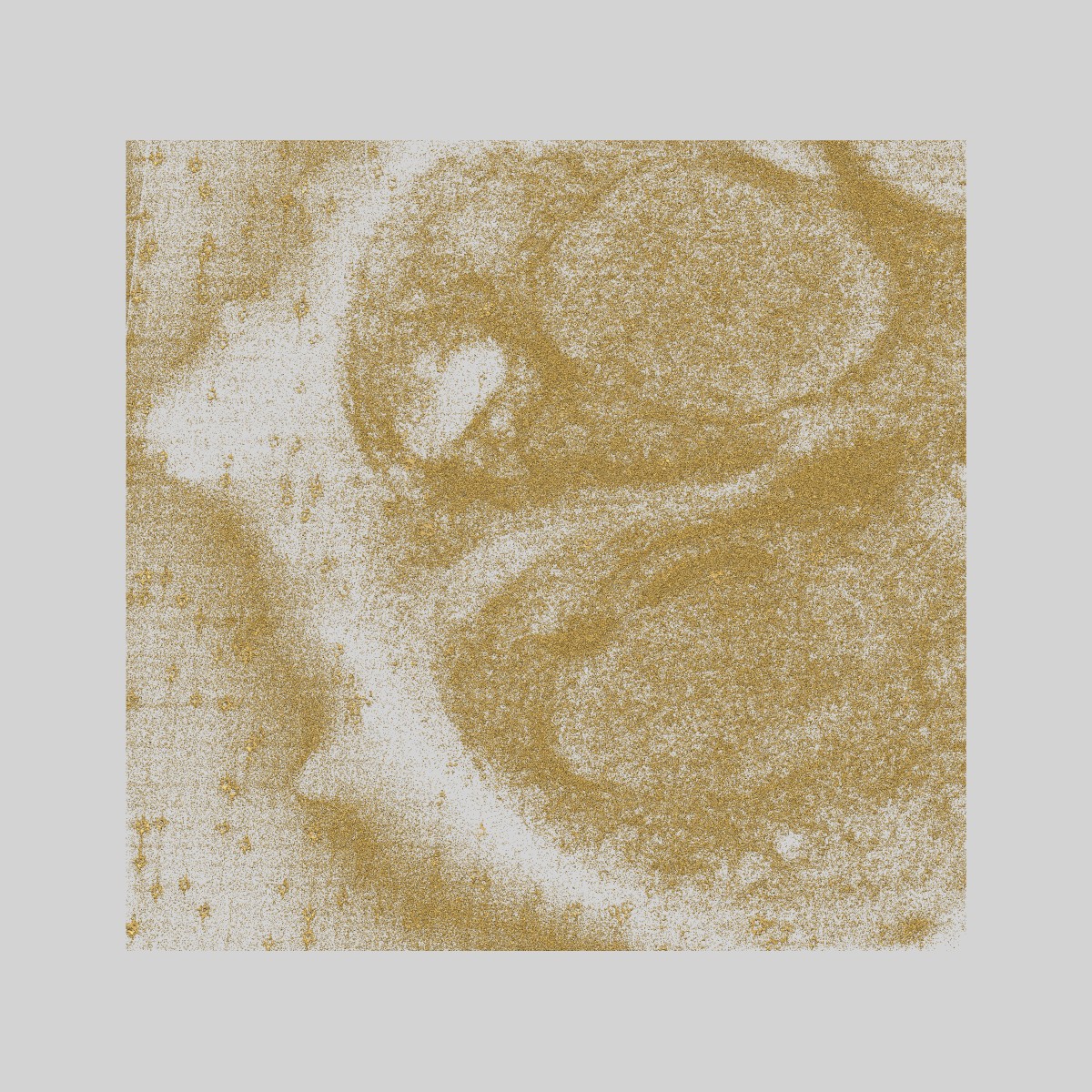
Malaria-infected red blood cell section showing partially segmented schizont
EMD-3586
Tomography Deposition: 10/02/2017
Map released: 22/03/2017
Last modified: 02/08/2017
Sample Organism:
Plasmodium falciparum 3D7
Sample: Plasmodium falciparum infected human erythrocyte treated with the egress inhibitor Compound 1
Raw data: EMPIAR-10087
Deposition Authors: Hale VL, Saibil HR
Sample: Plasmodium falciparum infected human erythrocyte treated with the egress inhibitor Compound 1
Raw data: EMPIAR-10087
Deposition Authors: Hale VL, Saibil HR
Parasitophorous vacuole poration precedes its rupture and rapid host erythrocyte cytoskeleton collapse in Plasmodium falciparum egress.
Hale VL  ,
Watermeyer JM,
Hackett F,
Vizcay-Barrena G,
van Ooij C,
Thomas JA ,
Spink MC,
Harkiolaki M,
Duke E,
Fleck RA ,
Blackman MJ,
Saibil HR
,
Watermeyer JM,
Hackett F,
Vizcay-Barrena G,
van Ooij C,
Thomas JA ,
Spink MC,
Harkiolaki M,
Duke E,
Fleck RA ,
Blackman MJ,
Saibil HR
(2017) PNAS , 114 , 3439 - 3444
 ,
Watermeyer JM,
Hackett F,
Vizcay-Barrena G,
van Ooij C,
Thomas JA ,
Spink MC,
Harkiolaki M,
Duke E,
Fleck RA ,
Blackman MJ,
Saibil HR
,
Watermeyer JM,
Hackett F,
Vizcay-Barrena G,
van Ooij C,
Thomas JA ,
Spink MC,
Harkiolaki M,
Duke E,
Fleck RA ,
Blackman MJ,
Saibil HR
(2017) PNAS , 114 , 3439 - 3444
Abstract:
In the asexual blood stages of malarial infection, merozoites invade erythrocytes and replicate within a parasitophorous vacuole to form daughter cells that eventually exit (egress) by sequential rupture of the vacuole and erythrocyte membranes. The current model is that PKG, a malarial cGMP-dependent protein kinase, triggers egress, activating malarial proteases and other effectors. Using selective inhibitors of either PKG or cysteine proteases to separately inhibit the sequential steps in membrane perforation, combined with video microscopy, electron tomography, electron energy loss spectroscopy, and soft X-ray tomography of mature intracellular Plasmodium falciparum parasites, we resolve intermediate steps in egress. We show that the parasitophorous vacuole membrane (PVM) is permeabilized 10-30 min before its PKG-triggered breakdown into multilayered vesicles. Just before PVM breakdown, the host red cell undergoes an abrupt, dramatic shape change due to the sudden breakdown of the erythrocyte cytoskeleton, before permeabilization and eventual rupture of the erythrocyte membrane to release the parasites. In contrast to the previous view of PKG-triggered initiation of egress and a gradual dismantling of the host erythrocyte cytoskeleton over the course of schizont development, our findings identify an initial step in egress and show that host cell cytoskeleton breakdown is restricted to a narrow time window within the final stages of egress.
In the asexual blood stages of malarial infection, merozoites invade erythrocytes and replicate within a parasitophorous vacuole to form daughter cells that eventually exit (egress) by sequential rupture of the vacuole and erythrocyte membranes. The current model is that PKG, a malarial cGMP-dependent protein kinase, triggers egress, activating malarial proteases and other effectors. Using selective inhibitors of either PKG or cysteine proteases to separately inhibit the sequential steps in membrane perforation, combined with video microscopy, electron tomography, electron energy loss spectroscopy, and soft X-ray tomography of mature intracellular Plasmodium falciparum parasites, we resolve intermediate steps in egress. We show that the parasitophorous vacuole membrane (PVM) is permeabilized 10-30 min before its PKG-triggered breakdown into multilayered vesicles. Just before PVM breakdown, the host red cell undergoes an abrupt, dramatic shape change due to the sudden breakdown of the erythrocyte cytoskeleton, before permeabilization and eventual rupture of the erythrocyte membrane to release the parasites. In contrast to the previous view of PKG-triggered initiation of egress and a gradual dismantling of the host erythrocyte cytoskeleton over the course of schizont development, our findings identify an initial step in egress and show that host cell cytoskeleton breakdown is restricted to a narrow time window within the final stages of egress.