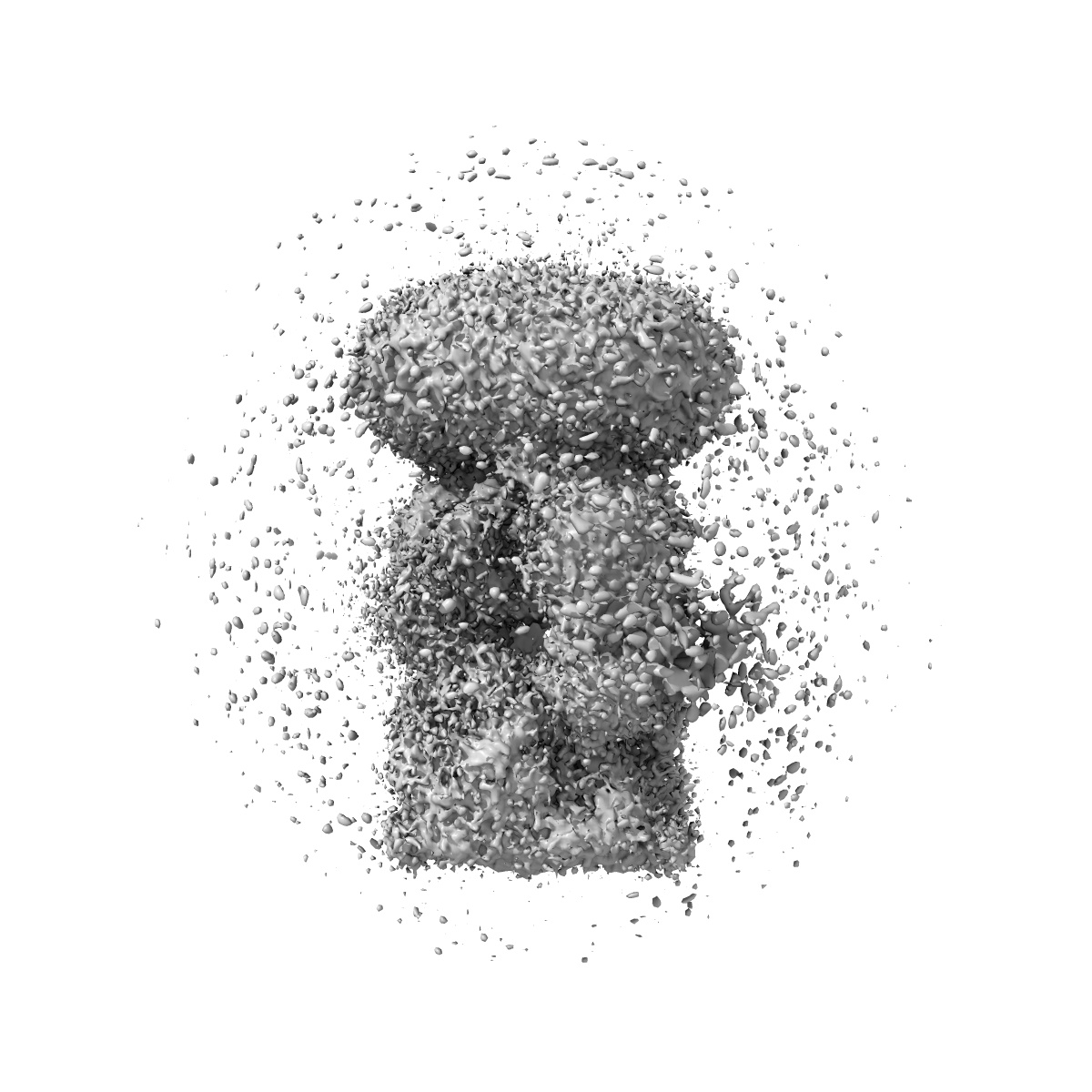
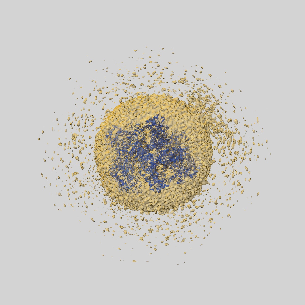
EMD-44130
Open state of kainate receptor GluK2 in complex with agonist glutamate and positive allosteric modulator BPAM344 bound to one concanavalin A dimer. Composite map.
EMD-44130
Composite mapSingle-particle
6.66 Å
 Deposition: 18/03/2024
Map released: 22/05/2024
Last modified: 13/11/2024
Sample Organism:
Rattus norvegicus,
Canavalia ensiformis
Sample: full-length rat GluK2 tetramer in complex with one concanavalin A homodimer
Fitted models: 9b37 (Avg. Q-score: 0.369)
Deposition Authors: Nadezhdin KD ,
Gangwar SP ,
Sobolevsky AI
,
Gangwar SP ,
Sobolevsky AI
Sample: full-length rat GluK2 tetramer in complex with one concanavalin A homodimer
Fitted models: 9b37 (Avg. Q-score: 0.369)
Deposition Authors: Nadezhdin KD
 ,
Gangwar SP ,
Sobolevsky AI
,
Gangwar SP ,
Sobolevsky AI
Kainate receptor channel opening and gating mechanism.
Gangwar SP ,
Yelshanskaya MV,
Nadezhdin KD ,
Yen LY,
Newton TP,
Aktolun M ,
Kurnikova MG ,
Sobolevsky AI
(2024) Nature , 630 , 762 - 768
,
Yelshanskaya MV,
Nadezhdin KD ,
Yen LY,
Newton TP,
Aktolun M ,
Kurnikova MG ,
Sobolevsky AI
(2024) Nature , 630 , 762 - 768
Abstract:
Kainate receptors, a subclass of ionotropic glutamate receptors, are tetrameric ligand-gated ion channels that mediate excitatory neurotransmission1-4. Kainate receptors modulate neuronal circuits and synaptic plasticity during the development and function of the central nervous system and are implicated in various neurological and psychiatric diseases, including epilepsy, depression, schizophrenia, anxiety and autism5-11. Although structures of kainate receptor domains and subunit assemblies are available12-18, the mechanism of kainate receptor gating remains poorly understood. Here we present cryo-electron microscopy structures of the kainate receptor GluK2 in the presence of the agonist glutamate and the positive allosteric modulators lectin concanavalin A and BPAM344. Concanavalin A and BPAM344 inhibit kainate receptor desensitization and prolong activation by acting as a spacer between the amino-terminal and ligand-binding domains and a stabilizer of the ligand-binding domain dimer interface, respectively. Channel opening involves the kinking of all four pore-forming M3 helices. Our structures reveal the molecular basis of kainate receptor gating, which could guide the development of drugs for treatment of neurological disorders.
Kainate receptors, a subclass of ionotropic glutamate receptors, are tetrameric ligand-gated ion channels that mediate excitatory neurotransmission1-4. Kainate receptors modulate neuronal circuits and synaptic plasticity during the development and function of the central nervous system and are implicated in various neurological and psychiatric diseases, including epilepsy, depression, schizophrenia, anxiety and autism5-11. Although structures of kainate receptor domains and subunit assemblies are available12-18, the mechanism of kainate receptor gating remains poorly understood. Here we present cryo-electron microscopy structures of the kainate receptor GluK2 in the presence of the agonist glutamate and the positive allosteric modulators lectin concanavalin A and BPAM344. Concanavalin A and BPAM344 inhibit kainate receptor desensitization and prolong activation by acting as a spacer between the amino-terminal and ligand-binding domains and a stabilizer of the ligand-binding domain dimer interface, respectively. Channel opening involves the kinking of all four pore-forming M3 helices. Our structures reveal the molecular basis of kainate receptor gating, which could guide the development of drugs for treatment of neurological disorders.