EMD-45674
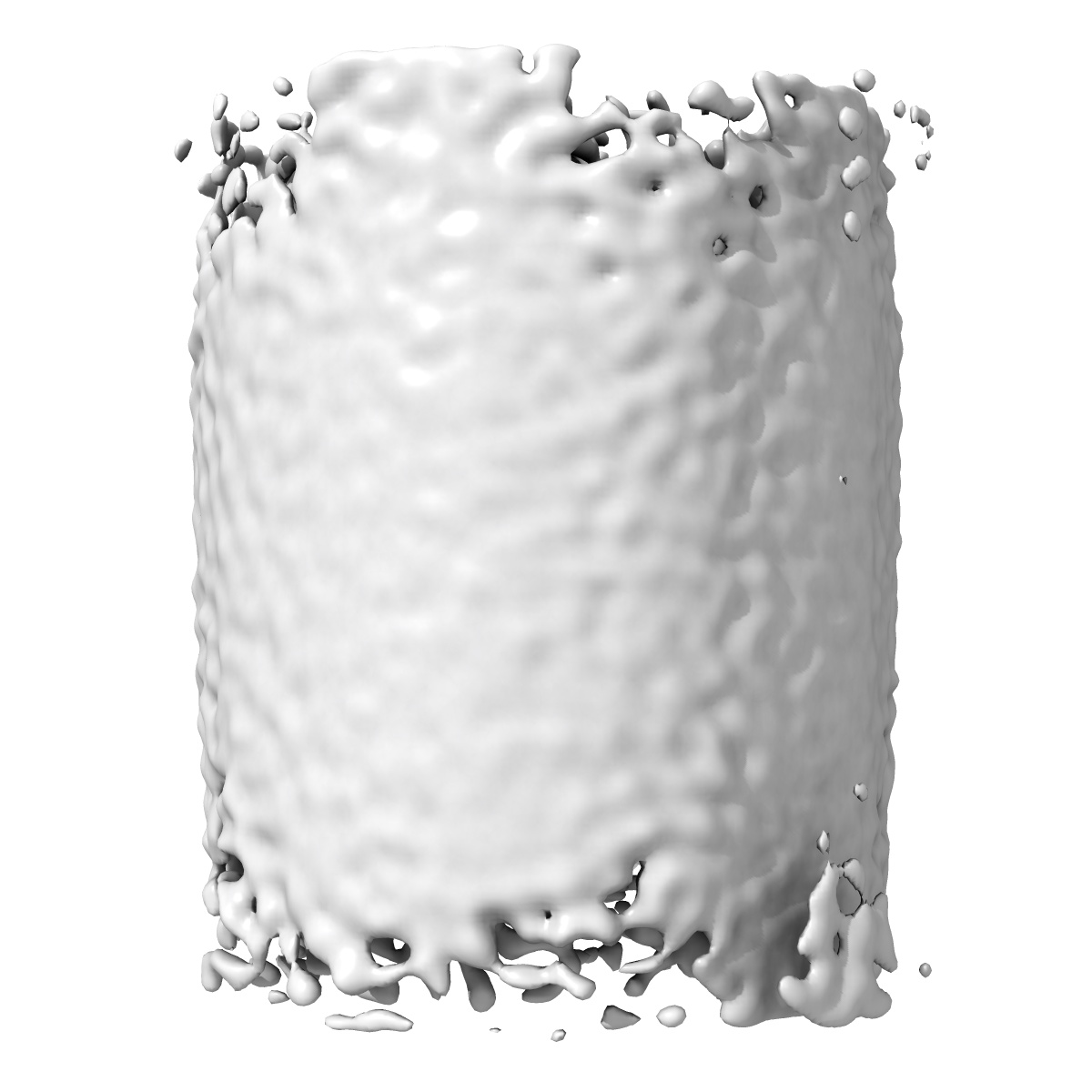
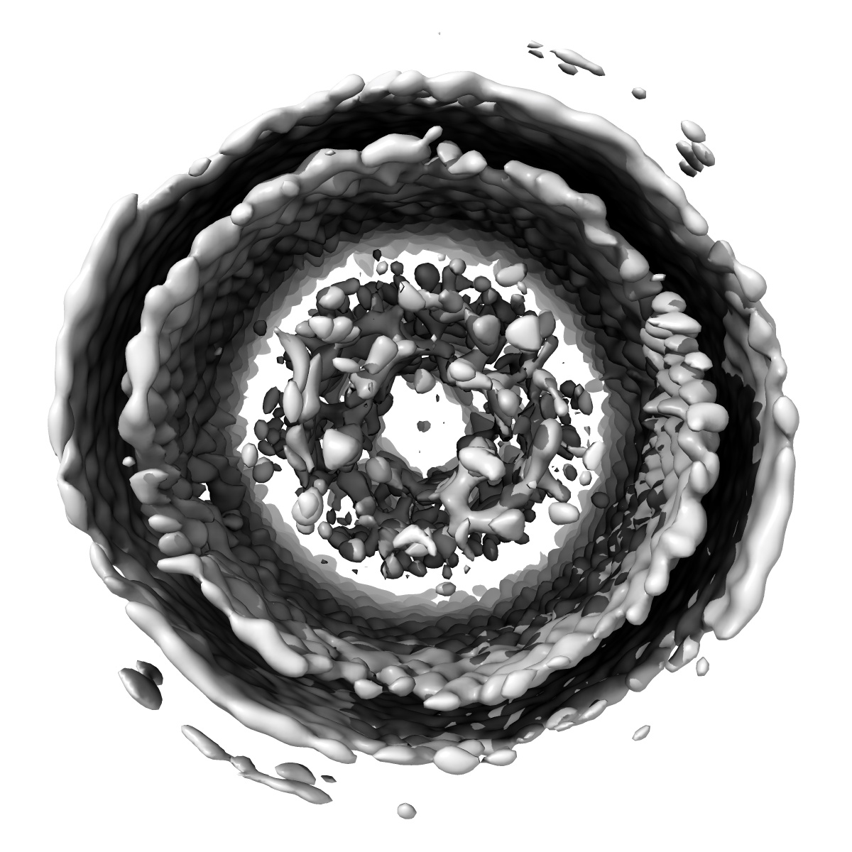
Subtomogram average of a whole Polar Tube cross-section from Encephalitozoon intestinalis microsporidian spores
EMD-45674
Subtomogram averaging70.0 Å
 Deposition: 10/07/2024
Map released: 26/02/2025
Last modified: 05/03/2025
Sample Organism:
Encephalitozoon intestinalis
Sample: Encephalitozoon intestinalis microsporidian spores
Deposition Authors: Usmani M ,
Coudray N ,
Bobe D ,
Kopylov M ,
Ekiert DC ,
Bhabha G
,
Coudray N ,
Bobe D ,
Kopylov M ,
Ekiert DC ,
Bhabha G
Sample: Encephalitozoon intestinalis microsporidian spores
Deposition Authors: Usmani M
 ,
Coudray N ,
Bobe D ,
Kopylov M ,
Ekiert DC ,
Bhabha G
,
Coudray N ,
Bobe D ,
Kopylov M ,
Ekiert DC ,
Bhabha G
Cryo-ET reveals the in situ architecture of the polar tube invasion apparatus from microsporidian parasites.
Usmani M ,
Coudray N ,
Riggi M ,
Raghu R ,
Ramchandani H,
Bobe D ,
Kopylov M ,
Zhong ED ,
Iwasa JH,
Ekiert DC ,
Bhabha G
(2024) bioRxiv
,
Coudray N ,
Riggi M ,
Raghu R ,
Ramchandani H,
Bobe D ,
Kopylov M ,
Zhong ED ,
Iwasa JH,
Ekiert DC ,
Bhabha G
(2024) bioRxiv
Grant Support:
- American Heart Association: 915749 (United States)
- Other private: Searle Scholars Program, SSP-2018-2737
- National Institutes of Health/National Institute Of Allergy and Infectious Diseases (NIH/NIAID): R01AI147131 (United States)
- Other private: Hirschl Career Scientist Award
- The Pew Charitable Trusts: PEW-00033055 (United States)
- Other government: NIH Common Fund Transformative High Resolution Cryo-Electron Microscopy, U24 GM129539
- Simons Foundation: SF349247 (United States)
- National Institutes of Health/National Institute of General Medical Sciences (NIH/NIGMS): 9 P41 GM103310 (United States)