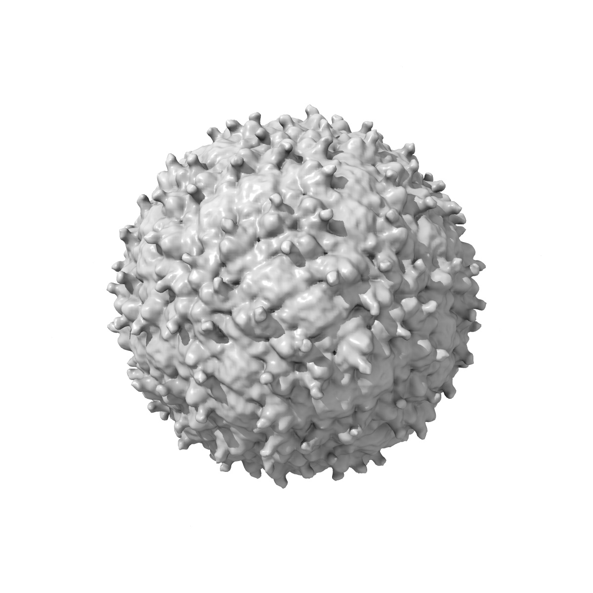
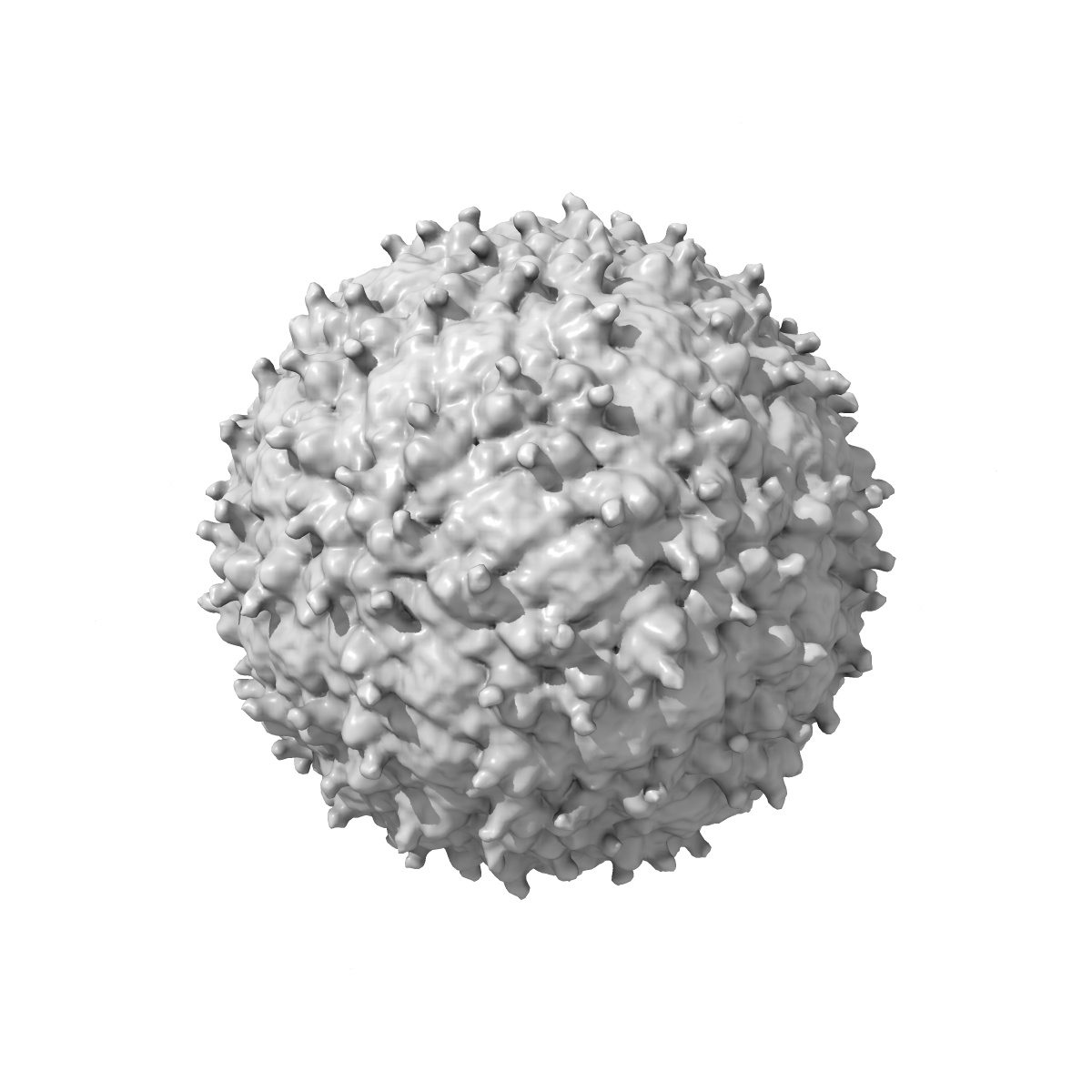
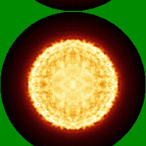
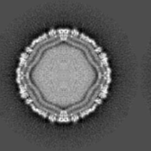
EMD-5296
cryo-EM reconstruction of West Nile virus
EMD-5296
Single-particle10.3 Å
 Deposition: 25/05/2011
Map released: 19/09/2012
Last modified: 24/07/2013
Sample Organism:
West Nile virus
Sample: West Nile Virus
Fitted models: 3j0b (Avg. Q-score: 0.086)
Deposition Authors: Zhang W ,
Kaufmann B,
Chipman PR ,
Kuhn RJ,
Rossmann MG
,
Kaufmann B,
Chipman PR ,
Kuhn RJ,
Rossmann MG
Sample: West Nile Virus
Fitted models: 3j0b (Avg. Q-score: 0.086)
Deposition Authors: Zhang W
 ,
Kaufmann B,
Chipman PR ,
Kuhn RJ,
Rossmann MG
,
Kaufmann B,
Chipman PR ,
Kuhn RJ,
Rossmann MG
Membrane curvature in flaviviruses.
Abstract:
Coordinated interplay between membrane proteins and the lipid bilayer is required for such processes as transporter function and the entrance of enveloped viruses into host cells. In this study, three-dimensional cryo-electron microscopy density maps of mature and immature flaviviruses were analyzed to assess the curvature of the membrane leaflets and its relation to membrane-bound viral glycoproteins. The overall morphology of the viral membrane is determined by the icosahedral scaffold composed of envelope (E) and membrane (M) proteins through interaction of the proteins' stem-anchor regions with the membrane. In localized regions, small membrane areas exhibit convex, concave, flat or saddle-shaped surfaces that are constrained by the specific protein organization within each membrane leaflet. These results suggest that the organization of membrane proteins in small enveloped viruses mediate the formation of membrane curvature.
Coordinated interplay between membrane proteins and the lipid bilayer is required for such processes as transporter function and the entrance of enveloped viruses into host cells. In this study, three-dimensional cryo-electron microscopy density maps of mature and immature flaviviruses were analyzed to assess the curvature of the membrane leaflets and its relation to membrane-bound viral glycoproteins. The overall morphology of the viral membrane is determined by the icosahedral scaffold composed of envelope (E) and membrane (M) proteins through interaction of the proteins' stem-anchor regions with the membrane. In localized regions, small membrane areas exhibit convex, concave, flat or saddle-shaped surfaces that are constrained by the specific protein organization within each membrane leaflet. These results suggest that the organization of membrane proteins in small enveloped viruses mediate the formation of membrane curvature.