EMD-5950
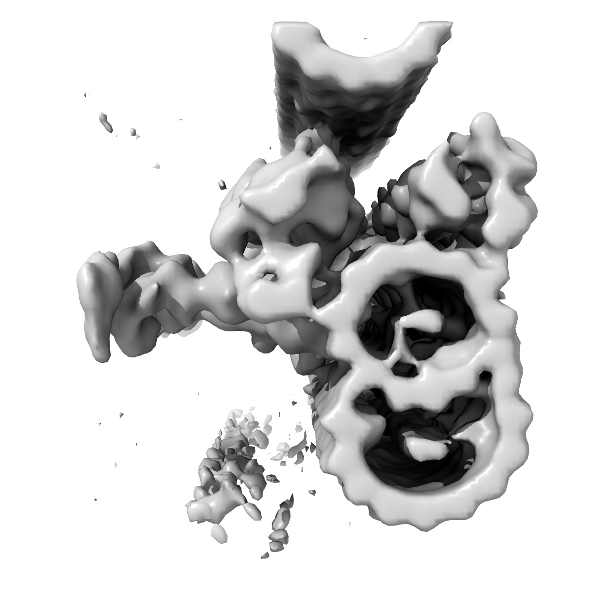
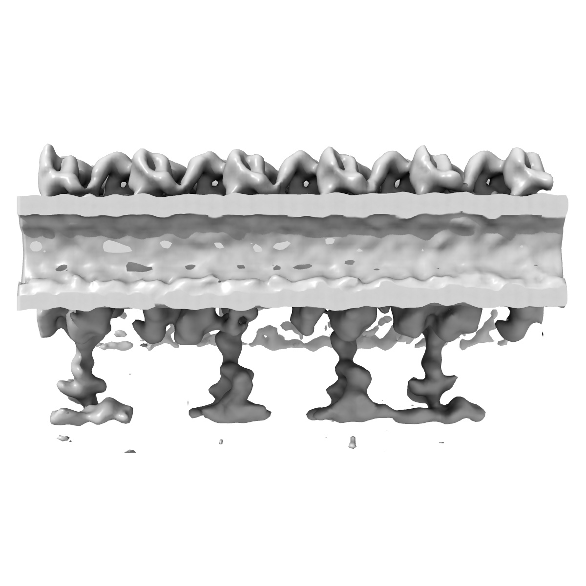
Cryo-electron tomography reveals ciliary defects underlying human RSPH1 primary ciliary dyskinesia
EMD-5950
Subtomogram averaging34.0 Å
 Deposition: 18/04/2014
Map released: 10/12/2014
Last modified: 17/12/2014
Sample Organism:
Homo sapiens
Sample: Normal human respiratory ciliary axonemes
Deposition Authors: Lin J ,
Yin W ,
Smith MC,
Song KK,
Leigh MW,
Zariwala MA,
Knowles MR,
Ostrowski LE,
Nicastro D
,
Yin W ,
Smith MC,
Song KK,
Leigh MW,
Zariwala MA,
Knowles MR,
Ostrowski LE,
Nicastro D
Sample: Normal human respiratory ciliary axonemes
Deposition Authors: Lin J
 ,
Yin W ,
Smith MC,
Song KK,
Leigh MW,
Zariwala MA,
Knowles MR,
Ostrowski LE,
Nicastro D
,
Yin W ,
Smith MC,
Song KK,
Leigh MW,
Zariwala MA,
Knowles MR,
Ostrowski LE,
Nicastro D
Cryo-electron tomography reveals ciliary defects underlying human RSPH1 primary ciliary dyskinesia.
Lin J ,
Yin W ,
Smith MC,
Song KK,
Leigh MW,
Zariwala MA,
Knowles MR,
Ostrowski LE,
Nicastro D
(2014) Nat Commun , 5 , 5727
,
Yin W ,
Smith MC,
Song KK,
Leigh MW,
Zariwala MA,
Knowles MR,
Ostrowski LE,
Nicastro D
(2014) Nat Commun , 5 , 5727
Abstract:
Cilia play essential roles in normal human development and health; cilia dysfunction results in diseases such as primary ciliary dyskinesia (PCD). Despite their importance, the native structure of human cilia is unknown, and structural defects in the cilia of patients are often undetectable or remain elusive because of heterogeneity. Here we develop an approach that enables visualization of human (patient) cilia at high-resolution using cryo-electron tomography of samples obtained noninvasively by nasal scrape biopsy. We present the native 3D structures of normal and PCD-causing RSPH1-mutant human respiratory cilia in unprecedented detail; this allows comparisons of cilia structure across evolutionarily distant species and reveals the previously unknown primary defect and the heterogeneous secondary defects in RSPH1-mutant cilia. Our data provide evidence for structural and functional heterogeneity in radial spokes, suggest a mechanism for the milder RSPH1 PCD phenotype and demonstrate that cryo-electron tomography can be applied to human disease by directly imaging patient samples.
Cilia play essential roles in normal human development and health; cilia dysfunction results in diseases such as primary ciliary dyskinesia (PCD). Despite their importance, the native structure of human cilia is unknown, and structural defects in the cilia of patients are often undetectable or remain elusive because of heterogeneity. Here we develop an approach that enables visualization of human (patient) cilia at high-resolution using cryo-electron tomography of samples obtained noninvasively by nasal scrape biopsy. We present the native 3D structures of normal and PCD-causing RSPH1-mutant human respiratory cilia in unprecedented detail; this allows comparisons of cilia structure across evolutionarily distant species and reveals the previously unknown primary defect and the heterogeneous secondary defects in RSPH1-mutant cilia. Our data provide evidence for structural and functional heterogeneity in radial spokes, suggest a mechanism for the milder RSPH1 PCD phenotype and demonstrate that cryo-electron tomography can be applied to human disease by directly imaging patient samples.