EMD-6447
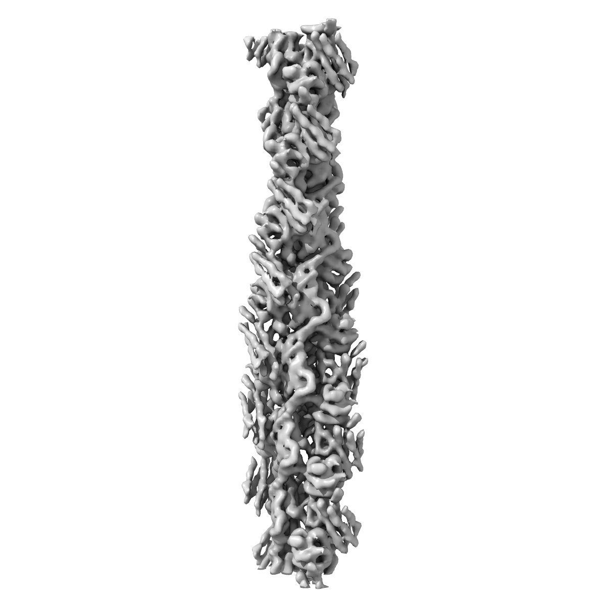
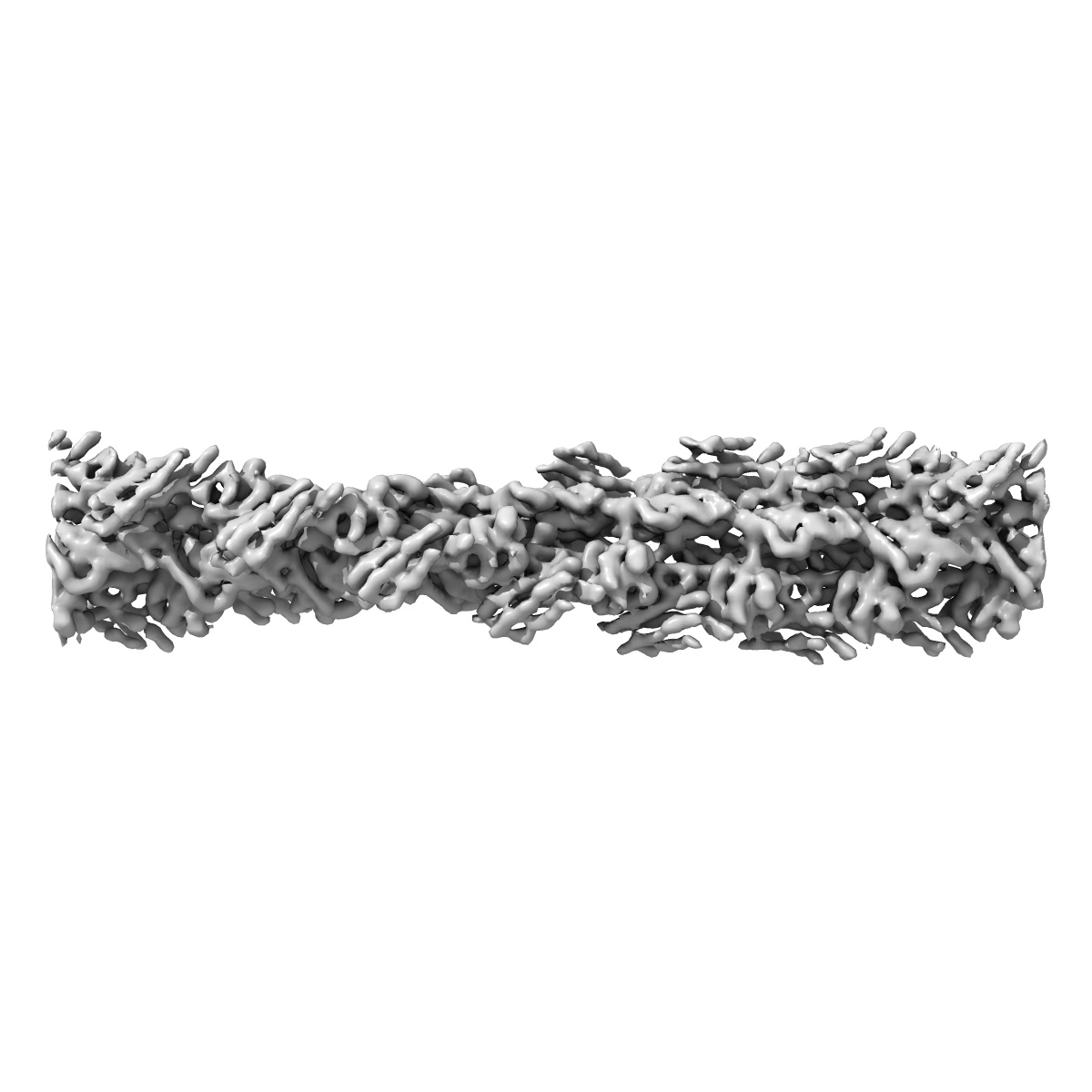
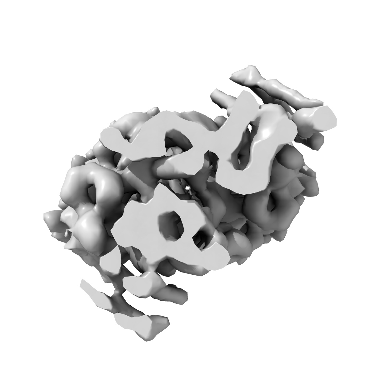
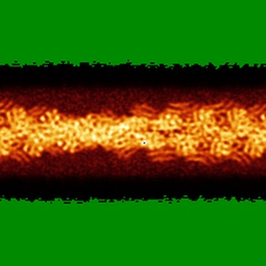
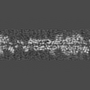
Cryo-EM reconstruction of the metavinculin-actin interface
EMD-6447
Helical reconstruction8.2 Å
 Deposition: 03/09/2015
Map released: 04/11/2015
Last modified: 17/02/2016
Sample Organism:
Oryctolagus cuniculus,
Homo sapiens
Sample: Metavinculin tail domain bound to F-actin
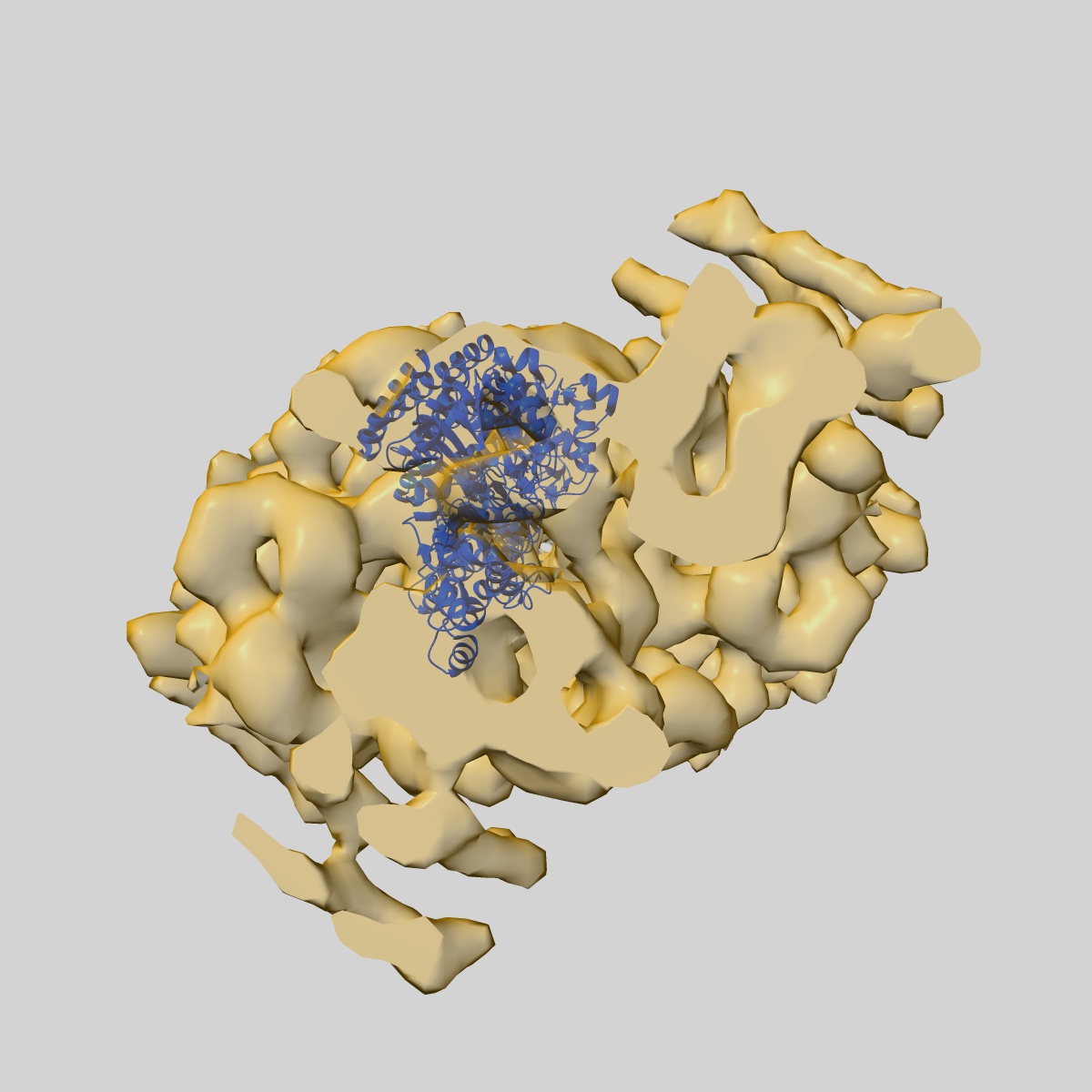
Fitted models: 3jbk (Avg. Q-score: 0.132)
Deposition Authors: Kim LY, Thompson PM, Lee HT, Pershad M, Campbell SL, Alushin GM
Sample: Metavinculin tail domain bound to F-actin
Fitted models: 3jbk (Avg. Q-score: 0.132)
Deposition Authors: Kim LY, Thompson PM, Lee HT, Pershad M, Campbell SL, Alushin GM
The structural basis of actin organization by vinculin and metavinculin.
Kim LY,
Thompson PM  ,
Lee HT ,
Pershad M,
Campbell SL ,
Alushin GM
,
Lee HT ,
Pershad M,
Campbell SL ,
Alushin GM
(2016) J. Mol. Biol. , 428 , 10 - 25
 ,
Lee HT ,
Pershad M,
Campbell SL ,
Alushin GM
,
Lee HT ,
Pershad M,
Campbell SL ,
Alushin GM
(2016) J. Mol. Biol. , 428 , 10 - 25
Abstract:
Vinculin is an essential adhesion protein that links membrane-bound integrin and cadherin receptors through their intracellular binding partners to filamentous actin, facilitating mechanotransduction. Here we present an 8.5-Å-resolution cryo-electron microscopy reconstruction and pseudo-atomic model of the vinculin tail (Vt) domain bound to F-actin. Upon actin engagement, the N-terminal "strap" and helix 1 are displaced from the Vt helical bundle to mediate actin bundling. We find that an analogous conformational change also occurs in the H1' helix of the tail domain of metavinculin (MVt) upon actin binding, a muscle-specific splice isoform that suppresses actin bundling by Vt. These data support a model in which metavinculin tunes the actin bundling activity of vinculin in a tissue-specific manner, providing a mechanistic framework for understanding metavinculin mutations associated with hereditary cardiomyopathies.
Vinculin is an essential adhesion protein that links membrane-bound integrin and cadherin receptors through their intracellular binding partners to filamentous actin, facilitating mechanotransduction. Here we present an 8.5-Å-resolution cryo-electron microscopy reconstruction and pseudo-atomic model of the vinculin tail (Vt) domain bound to F-actin. Upon actin engagement, the N-terminal "strap" and helix 1 are displaced from the Vt helical bundle to mediate actin bundling. We find that an analogous conformational change also occurs in the H1' helix of the tail domain of metavinculin (MVt) upon actin binding, a muscle-specific splice isoform that suppresses actin bundling by Vt. These data support a model in which metavinculin tunes the actin bundling activity of vinculin in a tissue-specific manner, providing a mechanistic framework for understanding metavinculin mutations associated with hereditary cardiomyopathies.