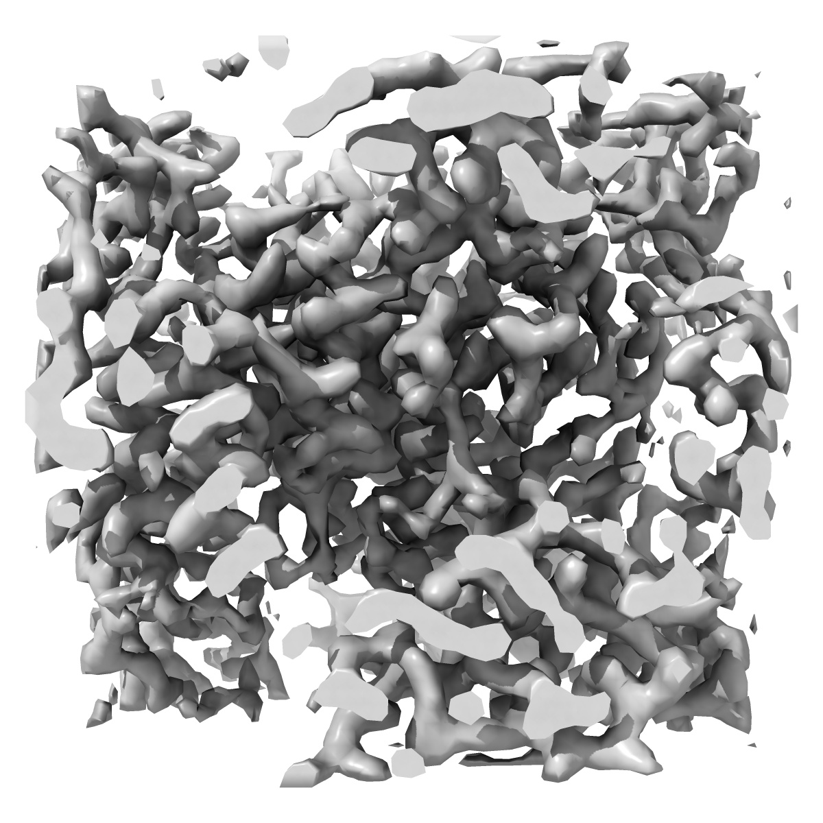
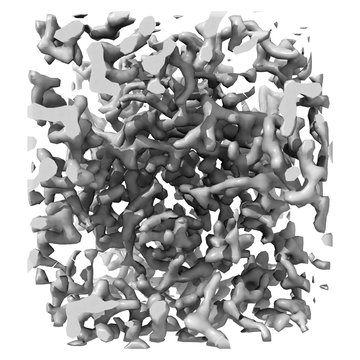
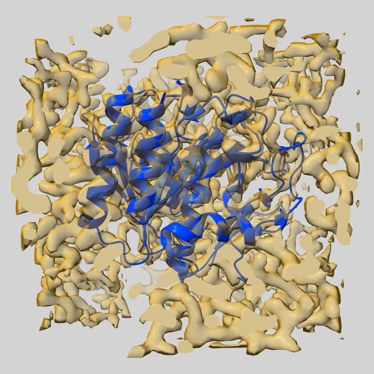
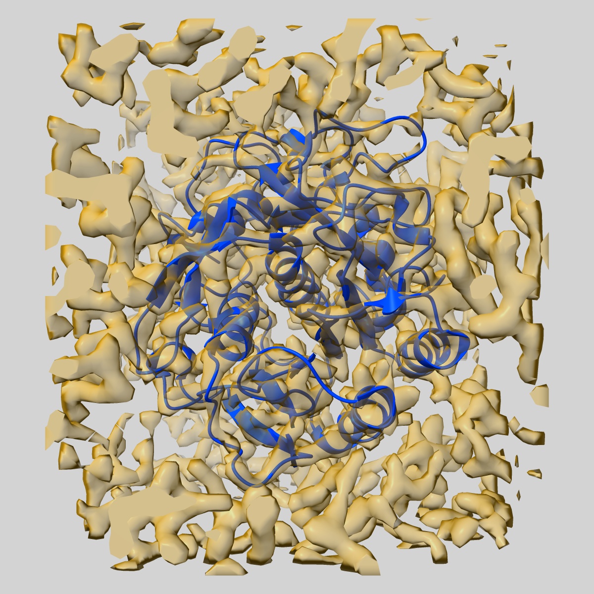
EMD-7494
3.20 A MicroED structure of proteinase K at 7.8 e- / A^2
EMD-7494
Electron Crystallography3.2 Å
 Deposition: 02/03/2018
Map released: 16/05/2018
Last modified: 20/11/2024
Sample Organism:
Parengyodontium album
Sample: Proteinase K
Fitted models: 6clb (Avg. Q-score: 0.601)
Deposition Authors: Hattne J ,
Shi D
,
Shi D
Sample: Proteinase K
Fitted models: 6clb (Avg. Q-score: 0.601)
Deposition Authors: Hattne J
 ,
Shi D
,
Shi D
Analysis of Global and Site-Specific Radiation Damage in Cryo-EM.
Hattne J ,
Shi D,
Glynn C,
Zee CT,
Gallagher-Jones M ,
Martynowycz MW,
Rodriguez JA,
Gonen T
(2018) Structure , 26 , 759 - 766.e4
,
Shi D,
Glynn C,
Zee CT,
Gallagher-Jones M ,
Martynowycz MW,
Rodriguez JA,
Gonen T
(2018) Structure , 26 , 759 - 766.e4
Abstract:
Micro-crystal electron diffraction (MicroED) combines the efficiency of electron scattering with diffraction to allow structure determination from nano-sized crystalline samples in cryoelectron microscopy (cryo-EM). It has been used to solve structures of a diverse set of biomolecules and materials, in some cases to sub-atomic resolution. However, little is known about the damaging effects of the electron beam on samples during such measurements. We assess global and site-specific damage from electron radiation on nanocrystals of proteinase K and of a prion hepta-peptide and find that the dynamics of electron-induced damage follow well-established trends observed in X-ray crystallography. Metal ions are perturbed, disulfide bonds are broken, and acidic side chains are decarboxylated while the diffracted intensities decay exponentially with increasing exposure. A better understanding of radiation damage in MicroED improves our assessment and processing of all types of cryo-EM data.
Micro-crystal electron diffraction (MicroED) combines the efficiency of electron scattering with diffraction to allow structure determination from nano-sized crystalline samples in cryoelectron microscopy (cryo-EM). It has been used to solve structures of a diverse set of biomolecules and materials, in some cases to sub-atomic resolution. However, little is known about the damaging effects of the electron beam on samples during such measurements. We assess global and site-specific damage from electron radiation on nanocrystals of proteinase K and of a prion hepta-peptide and find that the dynamics of electron-induced damage follow well-established trends observed in X-ray crystallography. Metal ions are perturbed, disulfide bonds are broken, and acidic side chains are decarboxylated while the diffracted intensities decay exponentially with increasing exposure. A better understanding of radiation damage in MicroED improves our assessment and processing of all types of cryo-EM data.