EMD-7822
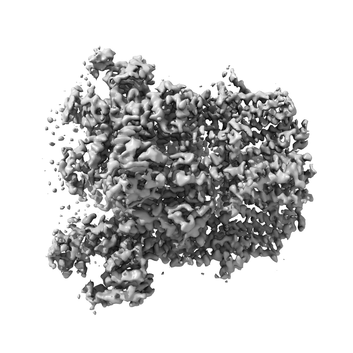
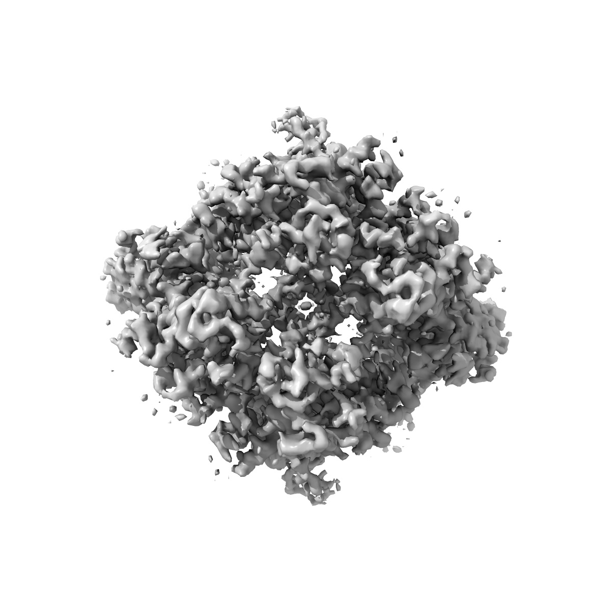
Cryo-EM structure of the zebrafish TRPM2 channel in the presence of Ca2+
EMD-7822
Single-particle3.8 Å
 Deposition: 23/04/2018
Map released: 15/05/2019
Last modified: 06/11/2024
Sample Organism:
Danio rerio
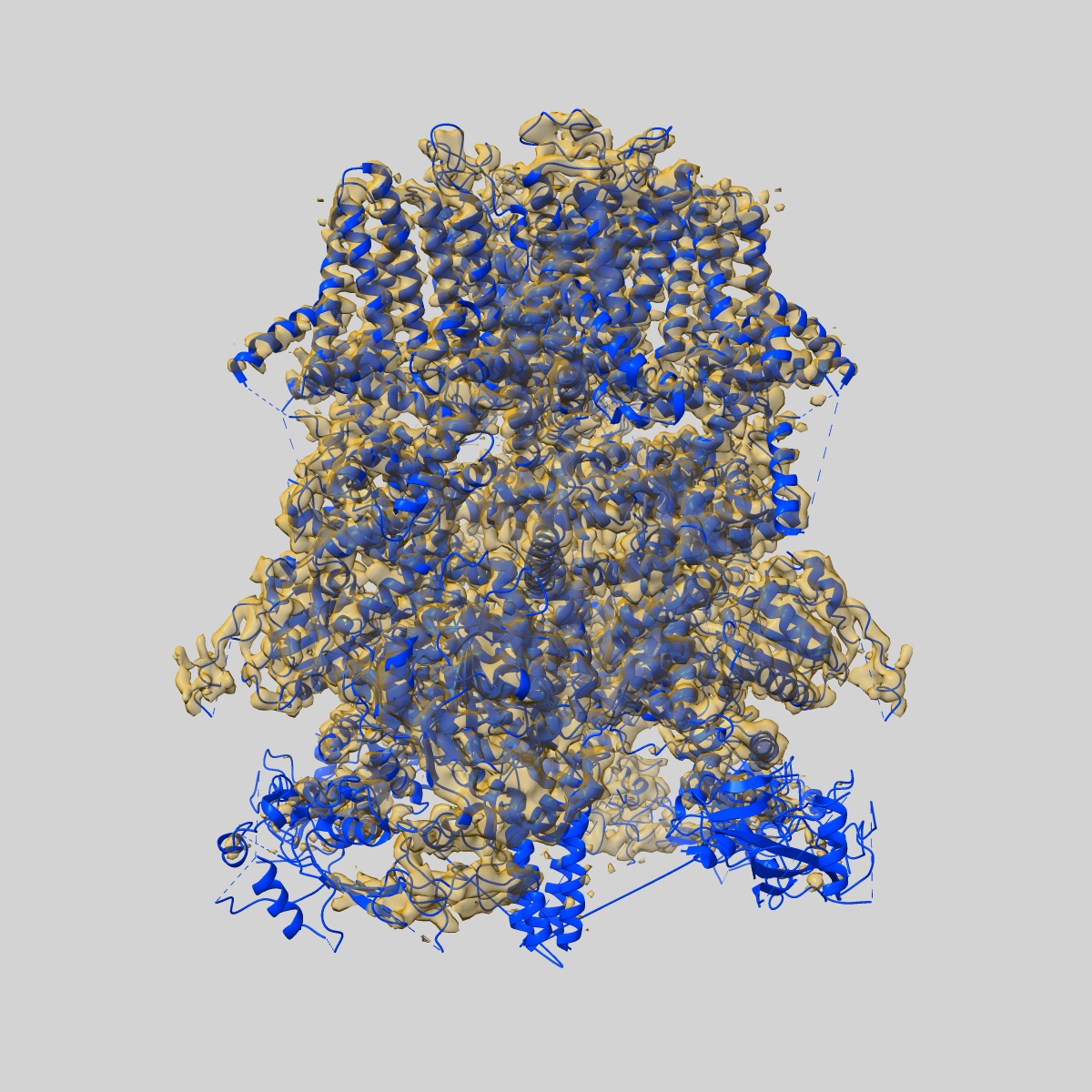
Sample: Transient receptor potential cation channel subfamily M member 2 (TRPM2)
Fitted models: 6d73 (Avg. Q-score: 0.365)
Deposition Authors: Yin Y ,
Wu M
,
Wu M
Sample: Transient receptor potential cation channel subfamily M member 2 (TRPM2)
Fitted models: 6d73 (Avg. Q-score: 0.365)
Deposition Authors: Yin Y
 ,
Wu M
,
Wu M
Visualizing structural transitions of ligand-dependent gating of the TRPM2 channel.
Yin Y ,
Wu M ,
Hsu AL ,
Borschel WF,
Borgnia MJ ,
Lander GC ,
Lee SY
(2019) Nat Commun , 10 , 3740 - 3740
,
Wu M ,
Hsu AL ,
Borschel WF,
Borgnia MJ ,
Lander GC ,
Lee SY
(2019) Nat Commun , 10 , 3740 - 3740
Abstract:
The transient receptor potential melastatin 2 (TRPM2) channel plays a key role in redox sensation in many cell types. Channel activation requires binding of both ADP-ribose (ADPR) and Ca2+. The recently published TRPM2 structures from Danio rerio in the ligand-free and the ADPR/Ca2+-bound conditions represent the channel in closed and open states, which uncovered substantial tertiary and quaternary conformational rearrangements. However, it is unclear how these rearrangements are achieved within the tetrameric channel during channel gating. Here we report the cryo-electron microscopy structures of Danio rerio TRPM2 in the absence of ligands, in complex with Ca2+ alone, and with both ADPR and Ca2+, resolved to ~4.3 Å, ~3.8 Å, and ~4.2 Å, respectively. In contrast to the published results, our studies capture ligand-bound TRPM2 structures in two-fold symmetric intermediate states, offering a glimpse of the structural transitions that bridge the closed and open conformations.
The transient receptor potential melastatin 2 (TRPM2) channel plays a key role in redox sensation in many cell types. Channel activation requires binding of both ADP-ribose (ADPR) and Ca2+. The recently published TRPM2 structures from Danio rerio in the ligand-free and the ADPR/Ca2+-bound conditions represent the channel in closed and open states, which uncovered substantial tertiary and quaternary conformational rearrangements. However, it is unclear how these rearrangements are achieved within the tetrameric channel during channel gating. Here we report the cryo-electron microscopy structures of Danio rerio TRPM2 in the absence of ligands, in complex with Ca2+ alone, and with both ADPR and Ca2+, resolved to ~4.3 Å, ~3.8 Å, and ~4.2 Å, respectively. In contrast to the published results, our studies capture ligand-bound TRPM2 structures in two-fold symmetric intermediate states, offering a glimpse of the structural transitions that bridge the closed and open conformations.